Thursday, November 25, 2021

Lupine Publishers | Open Access Journal of Oncology and Medicine (OAJOM)


Thanksgiving is a national holiday celebrated on various dates in the United States, Canada, Grenada, Saint Lucia, and Liberia. It began as a day of giving thanks and sacrifice for the blessing of the harvest and of the preceding year. Similarly named festival holidays occur in Germany and Japan. Thanksgiving is celebrated on the second Monday of October in Canada and on the fourth Thursday of November in the United States and around the same part of the year in other places. Although Thanksgiving has historical roots in religious and cultural traditions, it has long been celebrated as a secular holiday as well.

Monday, August 9, 2021

Lupine Publishers | Advances in Treating Relapsed Diffuse Large B Cell Lymphoma Treatment

 Lupine Publishers | Open Access Journal of Oncology and Medicine


Abstract

Diffuse large B cell lymphoma (DLBCL) is the most common type of non-Hodgkin lymphoma (NHL), comprising about 25% of all mature NHL. First-line therapy cures about 40-60% of patients. High dose chemotherapy followed by autologous stem cell transplant can cure about 50% of patients at relapse. Transplant-ineligible patients have shorter survival with every line of subsequent therapies with a median overall survival (OS) of 10 months at second line and 4.7 months at fourth lines of therapy. There is unmet need to treat patients with relapsed DLBCL. Chimeric antigen receptor T-Cells (CAR-T), with now three FDA- approved products to treat relapsed DLBCL, provide a cure in about 40% of patients. Other recently approved agents include antibody-drug conjugate targeting CD79b (Polatuzumab Vedotin), Anti-CD19 antibodies (Tafasitamab-cxix), and nuclear export inhibitor XPO1 (Selinexor) have provided a hope to patients with relapsed DLBCL. We will discuss these new approvals with comparison of response rate and side effect profiles.

Keywords: Diffuse Large B Cell Lymphoma; CAR-T Cell Therapy; Tafasitamab; Relapsed Refractory Lymphoma; Polatuzumab

Introduction


Non-Hodgkin lymphoma (NHL) is the eighth cause of cancer death. With an estimated 81,560 new cases and 20,720 deaths in 2021 [1] DLBCL is the most common type of NHL comprising about 25% of all NHL cases [2].

The prognosis depends on different factors at diagnosis including histological subtypes (Germinal center (GC) type versus non-GC subtypes) [3], genetic subtypes (double HIT) [4], patient age, stage of the disease, extra nodal involvement, and elevated LDH (IPI scoring) 5. Other important prognostic factors can be assessed after starting therapy, includes response at interim PET scan (PET scan after 2-4 cycles of therapy)6. Initial therapy for DLBCL remained the same for all different prognostic factors and different risk group which is the famous combination of chemotherapy R-CHOP (rituximab, cyclophosphamide, doxorubicin hydrochloride, vincristine, and prednisolone) despite multiple attempts to try to find a better alternative [7-9]. Except for high grade DLBCL with c-Myc and BCL2 and/or BCL6 gene rearrangement which most centers comfortable with using R-EPOCH chemotherapy regimen [10]. With the stander of care R-CHOP, about 30% of diffuse large B cell lymphoma patients relapse within the first 5 years [11]. Poor response to initial therapy and, or very early relapse (within the first six months), or what is called refractory disease is one of the worse predictors of poor survival based on data from scholar-1 study [12].

Current stander of care at relapse including high dose chemotherapy followed by autologous stem cell transplant [13]. In patient with very high-risk relapse (relapse within 1 year of therapy or refractory patients) there is a suggestion that early therapy with CAR-T cell may improve outcome. this question will be answered once we receive data from the now completed ZUMA7, and BELINDA and TRANSFORM trials comparing different CAR-T cell products (axicabtagene, Tisagenlecleucel, Lisocabtagene respectively) to the current stander of care i.e., high dose chemotherapy followed by autologous stem cell transplant in this very high-risk population. Treatment after relapse from second line of therapy inpatient who continue to maintain reasonable performance status is CAR-T cell therapy. Different CAR-T cell products are currently FDA approved to treat relapsed DLBCL after failure of at least two lines of therapy including Axicabtagene ciloleucel (Axi-Cel), Tisagenlecleucel and Lisocabtagene maraleucel(liso-cel). CAR-T provide about 40-55% complete response rate (CR). Most worrisome Toxicity including cytokine release syndrome (CRS) and neurological toxicity [14-16]. See Table 1 for comparing of different CAR-T cell products. Of note patient with DHL and DEL benefited from CAR-T cell therapy at relapse with Best overall response (ORR) was 56% (10/18) for DEL patients, 50% (5/10) for DHL patients which was not different from non-DHL/DEL in a retrospective data evaluation4. In patient who are intolerable to transplant or are ineligible for CAR-T therapy or had a relapse after CAR-T cell therapy there is no stander approach. Recent advances in newer agent gave some hope in managing these patients with improving survival.

Polatuzumab Vedotin

Polatuzumab Vedotin (Pola) is an antibody-drug conjugated that bind to CD79b which is present in all B-lymphocytes including mature malignant B-Cells delivering the microtubule inhibitor (MMAE) causing direct cytotoxicity. Polatuzumab alone or in combination with anti-CD20 antibody had a modest response with CR ranging from 0-13% [17-18]. The combination of Polatuzumab, Bendamustine and Rituximab was approved by FDA for patients with relapsed DLBCL after failure of at least 2 lines of therapy. In phase II randomized study comparing Pola BR to BR alone. The median age of patient on Pola BR arm was 67, 25% had prior stem cell transplant and 75% were refractory to prior line of therapy. ORR was 63% (25 of 40 patients) with CR rate of 40%. 48% of patients remained in CR at 1 year. Common side effects included fatigue, infusion reaction, neuropathy, and pancytopenia and increase risk of infection [19].

Tafasitamab

Tafasitamab (Tafa) is a Fc-enhanced, humanized, anti-CD19 monoclonal antibody. its FC portion is enhanced by modulating two amino acids that leads to increase affinity to Fcy receptors. As single agent Tafa has a response in DLBCL of 26% with responses lasting >12 months [20]. Phase II L-MIND study studied the combination of Tafa with lenalidomide as previous in vitro studies showed synergetic activity [21]. The study showed impressive CR rate of 43% with median duration of response of about 34 months. To compare the data from Pola-BR and Tafa-Lenalidomide combinations refer to (Table 2).

Selinexor

Selinexor is an XPO1(exportin 1) inhibitor. XPO1 is responsible for removal of multiple tumor suppressor genes out of the nucleus like P53, P73 and IkBk outside nucleus and inhabiting their function. The FDA had approved Selinexor as a single agent for the treatment of relapsed or refractory DLBCL after at least 2 lines of therapy. The approval was based on SADAL study, which was a Phase II study that included 134 patients, 45% were 70 years or older, 4% with DHL,41% had more than 3 lines of previous therapy with 72% refractory to the last line, 13% had elevated LDH. Single agent Selinexor resulted in ORR of 29%(95%CI:22,38) with CR rate of 13% with response duration of 6 months. Most common adverse reaction was fatigue, gastrointestinal side effects and pancytopenia [22]. Preclinical data suggest that the inhibition of XPO1 can provide a therapeutic target for DHL [23-24].

Bispecific T cell engager

Bispecific T-Cell engager (BiTe) therapy provide a promising off the shelve immune therapy for multiple cancers including NHL. In the contrary to CAR-T cell therapy, BiTe does not require manufacturing time or pre-infusion conditioning chemotherapy. Side effect can be similar but potentially less severe than CAR-T including CRS and neurological toxicity [25]. At this time BiTE therapy is not FDA approved to treat NHL but multiple trials across the world are testing different BiTe as single agent or with different combination including combining BiTe with Polatuzumab, with lenalidomide or other combinations. Recently published phase I/ II trial with Glofitamab, Bivalent CD20-target T-cell engaging BiTe. The study included 171 patients, 90% refectory to prior lines of therapy with median of 3 previous lines of therapy, 74% had DLBCL. ORR was in the phase II dose was 65.7% with CR of 57.1%. 84% of Cr patients had a maximum of 27.4 duration of response. Grade ¾ CRS was seen in 3.4% and grade 3 CNS toxicity in 1.2% [26]. Other promising BiTes including Blinatumomab, Epicoritamab, Monsunetuzumab and others [27-30].

Immune-Checkpoint Inhibitors in lymphoma

Immune check point inhibitors including programmed cell death /ligand inhibitors PD1 and PD-L1 inhibitors and CTL4 inhibitors have so far limited role in the therapy of DLBCL in the absence of biomarker targeted therapy. Future use may include pretesting for PD1 expression or the use of checkpoint inhibitors with combination [31].

CD47 Inhibitors

CD47 is present in virtually all cancer cells and it over expression is associated with poor prognosis. It is an inhibitory signal to macrophages (Do not Eat me signal) and inhabitation of CD47 lead to potentially increasing macrophage activation and tumor destruction. In addition, macrophages will present more tumor antigen and increase T-cell mediated cytotoxicity [32]. In Phase I/II trial combining CD47 inhibitor 5F9 with rituximab, the study included 22 patients (15 had DLBCL). Among patients with DLBCL, objective response rate was 40% with CR rate of 33%. Most common side effects were anemia and infusion reaction [33].

Conclusion and opinion

The treatment of relapsed DLBCL remains a challenge. With the approval of now three CAR-T cell therapy product we do have a chance of curing at least a third of those patients. Other may not benefit or may not have access to CAR-T cell therapy. Recently approved regimens that provide a good promise are the Tafa and lenalidomide combination and polatuzumab with Rituximab and Bendamustine combination. Although we have some patients with prolonged responses these combination does not provide a cure as of now. And most patients eventually succumb to their disease. With sequencing different combination and possibly prolonged maintenance therapy as, possible with the use of BiTE or other targeted agents we may see in the future an improved survival of relapsed refractory lymphoma.

Read More Lupine Publishers Oncology and medicine Articles : 


Read More Lupine Publishers blogger Articles : 
To Know more Open Access Publishers Click on Lupine Publisher:
Follow on Linkedin : https://www.linkedin.com/company/lupinepublishers
Follow on Twitter   :  https://twitter.com/lupine_online


Tuesday, May 26, 2020

Lupine Publishers | A Case Report and Review of Thymic Carcinoma with Adenoid Cystic Carcinoma like Features

Lupine Publishers | Open Access Journal of Oncology and Medicine


Introduction

Adenoid cystic carcinoma (ACC) is also called cylindroma, which is considered as a kind of low-grade malignant tumor and often occurs in head and neck salivary gland tissue. Most of them happened in submandibular gland and minor salivary glands while rarely happened in thymus. There are only 7 cases had already reported before all around the world. Our case is diagnosed as thymic carcinoma with adenoid cystic carcinomalike features by pathology after surgery; it is different with other cases because he had accepted a surgery six years ago because of the mediastinal mass and pleural effusion. Because of the symptom of chest tightness, he came to see doctor again and finally find there was a mediastinal mass and accepted the treatment of surgery. The medical history of our case maybe provides some inspiration of the pathogenic of thymic carcinoma with adenoid cystic carcinoma like features.

Information and history

A 60 years old male came to our hospital because of chest tightness in 2009/10. The enhanced chest CT showed “A large mass measuring 9.2*10.1cm² in the right side of the mediastinal which the average CT value is 9 Hu. The border with the superior vena cava and the right atrial and mediastinum is less clear. The right side of the chest is hydropneumothorax, Hydropericardium. The ultrasonic showed there is a mass of mixed echo in the right chest which had strong echo light and liquid visible mass dark space inside it. The test of pleural effusion by pleural puncture showed that was hematodes exudates without cancer cells. He accepted a surgery to resection of the mass by posterolateral thoracotomy, the pathology showed “Mostly considered as hematoma with organization according to clinical”. After the surgery ,he recovered and discharged from our hospital.
It is said by himself that there was found a mediastinal mass in his chest by CT in the year of 2010(the imaging data has been lost), and he taking chinese traditional medicein(herbal medicine) for tow month. After that he reexamination a chest CT showed the mediastinal mass was disappeared so he didn’t have the follow-up according to the doctor’s advice. 2014/10/13, he had a chest CT examination showed there is a 15*13cm² cystic with solid abnormal density shadow in the right front mediastium; Mediastinal lymph node is enlarged; Density of soft tissue in chest wall which is considered as metastatic. He reexamined a chest CT in 2015/1/4 but did not show significant change. Finally, he came to hospital again because of the intermittent chest pain. Complete the checks after hospitalization, lung function show” FEV1 : 3.94L, FEV1/FVC : 79%”, the enhanced chest CT showed “there is a 15.2*11.6cm² cystic with solid abnormal density shadow in the right front mediastium, the border is smooth and the density inside is uniform, the CT value is 18Hu; Right pulmonary atelectasis, the chest wall and diagram pars maybe have already metastasis tumor”.
a. Anamnesis: The patient with high blood pressure for more than 20 years, he use reserpin and Chinese traditional medicine to ctrl his high blood press by himself, but the effect is poor. His blood pressure was stabled for able 108/90mmHg for all years. Smoking 20 years and quit it for 23 years. We asked his medical history repeatedly, he denied the history of trauma and hemorrhagic disease.
b. Preoperative diagnosis: anterior mediastinum space occupying lesion, malignant tumor is the most possible. The chest wall maybe already metastasized. We suggested him to accept PET/CT to make the tumor’s metabolic strength clear and conform the primary focal; Or accept thoracentesis by fine needle to definite the pathological diagnosis. The patient and his family refused it and screamed for an operation.
So the surgery was planned. The patient was placed in the supine position under general anesthesia. Via median sternotomy. We saw the tumor was located in right anterior mediastinum, 12*12*10cm³, it was dense adhesion with mediastinal pleura, pericardium, diaphragm and right lung. The tumor is invading and partly wrapped around superior vena cava. Cut out part of the tumor tissue and sent to the examination of fast frozen section and the result was benign tumor. Then we completed removal of the tumor and cut one right rib and the tumor on the chest wall, the fast frozen section also report “benign tumor”. The tumor was solid and multicystic hemorrhagic with many sediment sample material. The diagnosis of the frozen section considered that most possible is encapsulated empyema. So we use polyninylpyrrolidone and normal saline to rinse the chest repeatedly and the closed thoracic conventionally.
The result of postoperative pathological: thymic carcinoma with adenoid cystic carcinomalike features with chest wall invasion, the tissue of cystiform is hyperplasia of fibrous tissue and stale hemorrhage. The result of immunohistochemical : CK(+), EMA(-), CK(L)(+), E-Ca(++), ER(+), PR(-), Ki67(10%), CK8/18(+), CD56 Focal(+), NSE(-), Syn(-), CgA(-).After the surgery, we checked the patient’s oral cavity, head and neck region but not found and obvious abnormal. 6 month pasted and we have not found any indication of tumor recurrence. He is still in follow up now.

Discussion


Adenoid cystic carcinoma is salivary gland tumor, but can still be reported in breast [1], lung [2], esophageal [3], postate [4]. Thymic carcinoma with adenoid cystic carcinoma like features is very rare. Only 7 cases have already be reported all around the world before while all cases is not in China.
Thymic carcinomas are defined as thymic epithelial neoplasmas, which are classified into 10 different histological types according to the 2015 update of the WHO classification, i.e.[5]
a. squamous-cell carcinoma
b. basal cell carcinoma
c. mucoepidermoid carcinoma
d. lymphoepithelioma-like carcinoma
e. sarcomatoid carcimoa
f. clear cell carcinoma
g. adenocarcinoma
h. NUT carcinoma
i. unclassified carcinoma
j. other rare thymic carcinoma.
Within the group of adenocarcinoma, four histologic subtypes are known: papillary adenocarcinoma; thymic carcinoma with adenoid cystic carcinomalikefeatures; mucinous adenocarcinoma; adenocarcinoma, non specifically. Compared with the 2004 update of the WHO classification, the name of adenoid cystic carcinoma is changed to thymic carcinoma with adenoid cystic carcinomalike features. It lack the real features of adenoid cystic carcinoma in the character of immunohistochemical because it is generate from the thymus (Table 1) [6-8].
Table 1: The character of the cases of thymic carcinoma with adenoid cystic carcinomalike features which are reported so far.
Lupinepublishers-openaccess-cancer-Oncology

The Character of Clinical

All of the clinical character of the cases which have already been reported is summarized above (chart-1). Thymic carcinoma with adenoid cystic carcinoma like features is very rare, most of them are occurred in elderly and few distant metastases. But it can show the character of aggressive growth and directly invasive chest wall; pericardium and other adjacent tissue. The initial symptoms of the patients are fever; cough; dyspnea; chest pain; the sense of suppression in the chest and the loss of weight [9]. thymic carcinoma with adenoid cystic carcinoma like features is generated in the tissue of thymus which located in the mediastinum, and the size of the most tumor is large. The severity of the clinical symptoms is uncorrelated with the size of the tumor.
Although the size of the tumor is large, the symptoms of patients is not severely even cannot let them go to see doctor. The clinical symptoms is formed because of the oppress by the tumor, there is no obviously symptoms in early stage, with the growth of tumor , the patient will appear the atypical symptoms such as cough; fever; dyspnea; chest pain and so on. Because thymic carcinoma with adenoid cystic carcinomalike features is a low grade malignancy, all cases had the features of gradual onset and grow progress. So only one case that had already metastases to rib was reported that is different whit the character of adenoid cystic carcinoma in salivary. According to the Masaoka stage of thymus tumor, our case is in stage β.

The Character of Pathology

The macroscopic view of the tumor: Most of them are consist of huge gritty and firm cystic solid mass with smooth border. Some of them have complete capsule. The cross-section showed multicystic, necrotic tissue mixed with fibrotic, yellow tissue nodules. Some of them can be observed the papillary hyperplasia and plenty of hemorrhagic effusion.
a. The microscopic view of the tumor: The character of our case is similar as salivary adenoid cystic carcinoma. The cystic and cribriform texture neoplastic nest is consist of basoloid cell carcinoma ,and bloody fluid or granular basophilic myxoid stroma full of the cyst. The microscopic view of the thymic carcinoma with adenoid cystic carcinomalike features can be classified as three types. If the tumor cell is undifferentiated, it will show the solid structure with irregular funicular or dense array by basaloid cells(solid type). If the tumor cell is differentiation to the glandular epithelium, it will show the gland tubular structure consist of the inner layer of columnar epithelium and the outside of the myoepithelial cells with homogeneous eosinophilic mucin full of the cavity of the tubule(tubular type). If the tumor cell is differentiation to the myoepithelial cell, it will show the cribriform structure with homogeneous basophilic cell matrix(cribriform type)[10]. The classify of thymic carcinoma with ACC like features is closely connected with the prognosis, so we infer that the prognosis of solid type is the worst while the prognosis of tubular type and the cribriform type is more better.
b. Immuno histochemistry: CK and CKL all showed positive in our case, they were marked at the epithelium which consist of cribriform structure. E-Ca(++) prompted the tumor has a certain invasive in accordance with the invasion of chest wall. Ki67(10%) prompted the active proliferation. CD56(focal +) is the new character that has not be reported before ER(+) prompted this case may be sensitive to endocrine therapy, but there isn’t any evidence of evidence-based medicine support it so the clinical significance needs to be confirmed.
Summarize other documents , we can get the following conclusion:
i. These marks is contribute to the diagnosis of thymic carcinoma with adenoid cystic carcinomalike features if they are positive: CollagenIV, laminin, P63, CK34betaE12, Ki67(1- 10%)
ii. These marks is contribute to the diagnosis of thymic carcinoma with adenoid cystic carcinomalike features if they are negative: Syn, NSE, CD117, CgA.
The molecular marks research of this disease has not yet been carried out. There are reports that suggested MYB-NFIB fusion gene, NF-κB, MMP-2, survivin is related to adenoid cystic carcinoma [11], and it is helpful to the molecular marks research of thymic carcinoma with adenoid cystic carcinomalike features.

The Differential Diagnosis

PET/CT has great significance to this disease in differential diagnosis, it can estimate whether the mass is primary tumor or metastases. The report suggested the tumor of this disease show high value of SUV max in PET/CT while its metastases can also show unusually high value of SUV max [9]. The retrospective analysis show that have a examination of PET /CT before surgery is correct and meaningful. In the primary thymic carcinoma, this disease should be distinguished with basaloma. In morphology, most of the tumor cell of basaloma is cubic cells with small and deep dyeing nucleus which array form lobulated or funicular [12,13]. The results of Immuno histochemistry can confirm that our case can be diagnosed as thymic carcinoma with adenoid cystic carcinoma like features.

The treatment and prognosis

Surgery is given priority to this disease at present and radiation therapy can according to the situation. The effect of chemotherapy for this disease is poor and if take chemotherapy as a routine treatment is still controversial. Some professional insist of that adenoid cyst carcinoma cannot get enough benefit from chemotherapy, so we should treat chemotherapy as the last treatment [14]. We believe that A case report and review of thymic carcinoma with adenoid cystic carcinoma like features is a low level malignant tumor so the it should have better prognosis after standardized treatment. But we still should be carefully evaluated before surgery and if we suspect the diagnosis as thymic tumor with ACC like carcinoma or huge mediastinal mass, PET/CT will be suggested to confirm the quality of the mass and whether there is metastases or not.
Locally metastasis and the chest wall invasion should not be the absolute contraindications of surgery because as a low level malignant tumor, if we resection the tumor and metastases completely and take some radiation therapy appropriately, nice quality of life will get. In our case a recurrence is appear after the surgery in 2009, but he said itself that the mediastinal mass was once disappeared after the treatment of Chinese traditional medicine. So we guess that Chinese traditional medicine may effective for this disease.

The discussion of pathogenesis

A case report and review of thymic carcinoma with adenoid cystic Carcinoma like features is a kind of thymic carcinoma, it is generated from thymic epithelial cells. If the tumor cell is differentiation to the myoepithelial cell, it will show the cribriform structure with homogeneous basophilic cell matrix. The medical history of our case is so specially that he found the mediastinal mass six years ago and experienced the surgery. The pathological report at the time of 2009 showed “hematoma with organization”. Organization means the absorption process by new granulation tissue if necrotic tissue, thrombus, .com or foreign body cannot be dissolved or absorbed or separated or discharged. The granulation tissue contains abundant myocyte and some of them will differentiate to myofibroblast after the stimulate of cytokines.
And these cytokines that can induce differentiation may induce the differentiate thymic epithelial cell to myoepithelium and finally generate to the type of cribriform of thymic carcinoma with adenoid cystic carcinomalike features is a kind of thymic carcinoma. This maybe one of the triggers of this disease although still need to establish by experiments. According to the inference above, we consider coagulate hemothorax and mediastinal hematoram may result thymic carcinoma with adenoid cystic carcinoma like features. In order to avoid the remained thymus tissue be stimulated to differentiate to thymic carcinoma, in the surgery of mediastinal hematoma, thymectomy completely is necessary and fat tissue of mediastinal should be resection completely too.

Read More Lupine Publishers Oncology and medicine Articles : 

Read More Lupine Publishers blogger Articles : 
To Know more Open Access Publishers Click on Lupine Publisher:
Follow on Linkedin : https://www.linkedin.com/company/lupinepublishers
Follow on Twitter   :  https://twitter.com/lupine_online 


Wednesday, May 13, 2020

Lupine Publishers | Promising Role of Fractional Calculus in Biomedicine and Biophysics

Lupine Publishers | Open Access Journal of Oncology and Medicine


Introduction
The study of complex systems and investigation of their structural and dynamical properties have attracted considerable interests among scientists in general and physicists, biologists and medical researchers in particular. Complex systems can be found almost everywhere however the highest level of complexities is related to living and biological organisms and systems. Due to the lack of a reliable and effective tool to investigate such systems, we have not reached to the complete understanding and comprehensive pictures of the phenomena and processes which occur in these systems. Of course a comprehensive knowledge of biological and biomedical complex phenomena will be achieved when we employ simultaneously different field of science and engineering including: biology, chemistry, physics, mathematics, mechanical engineering and so on.
Fortunately in recent year's powerful tool of fractional calculus has been proposed for study of complex and nonlinear phenomena. It is in fact very useful tool for describing the behavior of nonlinear systems which are characterized by: special kind of non-locality, long-term memory and fractal properties. There exist many biological objects and systems with memory, nonlocal effects and nonlinear behaviors and such these non-localities and memory effects in biological objects and systems mean that the next state of the organism or system relies not only on its present state but also upon all of its previous states. As a result, the concept of fractional dynamics and in fact adopting fractional calculus can play an important role in the study of dynamical biological systems. Up to now few number of important issues such as: protein folding phenomena and mechanics of cancer cells (for more details see the references which have investigated physics of protein and physics of cancer in detail) have been investigated using the framework of fractional dynamics [1].
However many other important issues still remain as open issues, such as: modeling of interactions between light (laser) and biological tissue and modeling of intracellular (and intercellular) interactions in the framework of fractional dynamics. As a physicist or biologists and even medical researchers, we always are able to model natural phenomena for instance modeling of tumor growth using systems of differential equations and nowadays it is well know that the fractional-order ones are more comprehensive and also incorporate memory effect and the concept of non-locality in the model.
Mathematically the idea is in fact, to rewrite the ordinary governing differential equations in the fractional form by replacing the standard derivative with a fractional derivative of arbitrary order which is defined in the Caputo sense as follows:
where Γ denotes the Gamma function and , . And its Laplace transform can be given by:

Where, F(s) is the Laplace transform of f (t). Solutions of fractional differential equations generally will be expressed using a generalized special function named as Mittag-Leffler function. This function can be considered as a generalized exponential function and has several different forms. For instance the one-parameter Mittag-Leffler function is defined by the series expansion as:
Where 
C is the set of complex numbers? It is worth mentioning that the exponential function is just a special case of Î± = l Mittag-Leffler function, for example for the special case of , the Mittag-Leffler function Eq. (3) reduces to the exponential function E
1(z) = ez . This point is very important because of that the natural exponential function has been considered as a fundamental function of natural science and in particular biology up to now, so that many phenomena could be described using it and now scientist are able to think that with such this new framework (i.e. fractional differential equations and their solutions in terms of Mittag-Leffler functions) they can find many new results and information about biological and biomedical phenomena [2,3].
Finally, based on all above mentioned reasons, as a conclusion we should say that we believe that the powerful tool of fractional calculus and in fact the frame work of fractional dynamics can give.com new insights in understanding and modeling of nonlinear complex phenomena in various living cellular structures and their interactions and we invite all biologist and medical researchers to consider this new powerful approach for their future studies.

Read More Lupine Publishers Oncology and medicine Articles : https://lupine-publishers-cancer-journal.blogspot.com/

Read More Lupine Publishers blogger Articles : https://lupinepublishers.blogspot.com

To Know more Open Access Publishers Click on Lupine Publisher

Follow on Twitter   :  https://twitter.com/lupine_online  

Thursday, March 12, 2020

Lupine Publishers: Lupine Publishers | Principles of the Military Con...

Lupine Publishers: Lupine Publishers | Principles of the Military Con...: Lupine Publishers- Anthropological and Archaeological Sciences Journal Impact Factor   Introduction A way from the regular th...

Lupine Publishers | Acute Erythroblastic Leukemia Revealed by Dermatological Manifestations

Lupine Publishers | Open Access Journal of Oncology and Medicine

Abstract

Acute erythroblastic leukemia is characterized by the proliferation of a predominant erythrocyte population on other lineages. Cutaneous manifestations remain rare and misleading, making the diagnosis of difficult to suspect as first-line. Here, we report an unusual and rare case of acute leukemia in a 24 year old male with gingival hypertrophy and dermatological manifestations. This case emphasizes that dentist and dermatologist should be well acquainted with these manifestations of systemic diseases.

Case Report

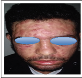
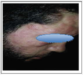
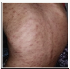
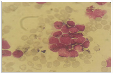
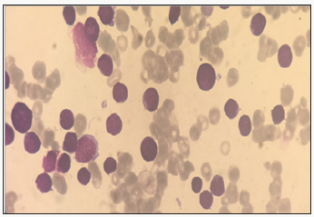
We report the case of 24 years old patient, with no significant pathological history, who had a rash for 10 days in a context of fever and very bad general condition. At admission the patient was febrile, tachycardic and dyspneic. Physical examination revealed erythemato-purplished papulo-nodules on the face, trunk, limbs and a gingival hyperplasia. The oral state was deplorable. Bilateral cervical lymphadenopathy was also found without the patosplenomegaly (Figures 1-3). The biological assessment showed a CRP of 150 and a pancytopenia with a Hbat 7.5g / dl, normal VGM and CCMH, a deep thrombocytopeniaat 85000 / l, leukocytesat 1500 / l. The blood smear showed 35% of circulating blasts and 22% of erythroblasts (Figure 4).
Figure 1: Clinical Manifestations of AML.
Figure 2: Clinical Manifestations of AML.
Figure 3: Clinical Manifestations of AML.
Figure 4: Blood smear showing 35% of circulating blasts.
The medullo gram showed a hyper-cellular marrow with a rate of myeloblasts greater than 45% compared to all non-erythroblastic elements and erythroblastic hyperplasia estimated at more than 65%; with signs of dyserythropoiesis suggestive of the diagnosis of erythroleukemia (Figure 5). Blood immune phenol typing was positive for CD13, CD33, MPO and Glycophotin A. The evolution was unfavourable; the patient died due to massive alveolarhemorrhage.
Figure 5: Hyper cellular marrow infiltrated by a blastic contingent estimated at 45%.

Discussion

Acute erythroblastic leukemia is characterized by the proliferation of a pre dominantery throcyte population on other lineages. There are two types: Erythroleukemia: defined by the presence in the bone marrow of more than 50% of the erythroid precursors of all the medullary cells, and more than 20% of myeloblasts of the whole non-erythrocytemedullary cells - Pure erythroid leukemia: it presents a neoplastic proliferation made of more than 80% of erythrocyte cells without obvious presence of the myeloblastic contingent [1]. It is usually manifested by signs of bone marrow failure and cytopenia [2,3], skin involvement remains rare, varied and disorienting the diagnosis; they are found mainly in Acute myelovlastic leukemia [4,5]. Cutaneous manifestations during leukemia are infrequent and varied. They designate all the cutaneouslesions related to the haematological malignancy directly or indirectly following their treatment; we essentially distinguish.
The specific dermatological lesions which can reveal hematological diseases [4], are mainly represented by leukaemides (leukaemia cutis), which are red brown to purple dermal papules, plaques or nodules. Granulocyticsarcom as an extra-medullary tumour masses, ulcerated plaques and gingival hypertrophy [5]. The infectious dermatoses secondary to the biological disturbances accompanying the malignan themopathy and their treatments. The occurrence of specific cutaneouslesions in leukemia is synonymous with a major aggravation of the prognosis (with for example a survival twice as short if there is a specific cutaneous involvement); this seriousness make some authors propose different treatments with a medium-long stay hospitalization[6-8].
In our case, acute myelonlastic leukemia 6 (AML 6 ) was revealed by diffuse leukemias resulting from the infiltration and proliferation of malignantha ematological cells (blasts) in the skin and by gingival hyperplasia secondary to mucosal infiltration [5]. The clinical presentation of acute leukemia including AML6 in the form of ulcer ativenecroticgingivitis in the foreground, is a rare form to be remembered, mentioned in all courses of medicine and dentistry, stipulating that Gingival involvement is a classic feature of leukemia [6] The frequent association of skin cancers with haematological malignancies is also highlighted in several publications [5].

Conclusion

The cutaneous localizations are among the rarest extreme dullary lesions of acute myeloid leukaemia’s (AML) not exceeding 1%. They are generally considered as factors of worse prognosis. Their cytogenetic or mutational specificities remain un established to date.


For More Lupine Publishers Open Access Journals Please visit our Website:

For more Open Access Journal of Oncology and Medicine Please Click Here: https://lupinepublishers.com/cancer-journal/

 
Follow on Twitter   :  https://twitter.com/lupine_online


Thursday, February 27, 2020

Lupine Publishers | Subjection between Breast Cancer and Body Mass Index, the Role of L-Carnitine in Prediction and Outcomes of the Disease

Lupine Publishers | Open Access Journal of Oncology and Medicine


Abstract


Increasing the effectiveness of antitumor therapy in breast cancer patients who take L-carnitine during preoperative systemic antitumor therapy compared with patients receiving standard neoadjuvant systemic antitumor therapy served as a prerequisite for studying possible antitumor mechanisms of L-carnitine. The positive effect of L-carnitine is due to the transfer of palm-n-LC through the inner membrane into the mitochondrial matrix, which promotes the formation of a significant number of ATP molecules. It has also been shown that L-carnitine can have a double protective effect, enhancing the energy dynamics of the cell and inhibiting the hyperexcitability of the cell membrane, that making it an ideal nutrient for the prevention and treatment of cancer. This article summarizes the results of epidemiological and clinical studies of the use of L-carnitine in the treatment of breast cancer
Keywords: Body mass index (BMI); Breast cancer (BC); Obesity; Overall survival; L carnitine


Introduction

The incidence of breast cancer in the world in general and in Ukraine in particular is growing. In 2017, in Ukraine the incidence reached 16 percent of female population, for which, the breast cancer ranked first in structure of oncological incidence among women. In analyzing the data of the National Cancer Registry of Ukraine, it should be noted, that in comparison with 2014 year, the prevalence rate of breast cancer in 2016has increased by 5,1%, that indicates importance of improvement diagnostic procedures and methods of treatment it [1]. Studying the scientific literature on this subject, we noticed that there is a strong biological relationship between obesity and a poor outcome of breast cancer. And having analysed the date of Ministry of Health in Ukraine it can be concluded, that about 26% of women in 2017 year had overweight or obesity.
Obesity has a chronic metabolic character, which is the result of the interaction of the endogenous factors, environmental conditions and lifestyle. Endogenous factors could be considered a violation of the genetic and hormonal balance. The external conditions and type of lifestyle include irregular rhythm nutrition, use of substandard products and sedentary lifestyle. Obesity is the first risk factor for metabolic syndrome, diabetes type II, cardiovascular disease and some forms of cancer, including breast cancer. Since overweight is a risk factor for breast cancer, there is reason to believe that among patients with breast cancer the percentage of obese women is higher than in the population. The risk of breast cancer in postmenopausal women by 30%, it is more than in premenopausal, women with obesity-50%. Furthermore it was proven that obesity is associated with poor prognosis in patients with breast cancer, regardless of menopausal status, and effectiveness of systemic medication breast cancer in patients that have over weight is lower than in patients with normal BMI.
Although obesity is associated with a poor outcome in women with breast cancer, it is unclear how weight loss after diagnosis will change its course and results. Recently, complementary and alternative medicine (CAM) is widely accepted among patients with breast cancer, which may provide several beneficial effects including reduction of therapy-associated toxicity, improvement of cancer-related symptoms, fostering of the immune system, and even direct anticancer effects [2]. L-carnitine is a metabolite of C4 oil LC, which is involved in the transfer of palm-n-LC through the inner membrane into the mitochondrial matrix and is a substrate for the formation of ATP molecules. Carnitine is a trim ethylated amino acid naturally synthesized in the liver, brain and kidneys from protein lysine and methionine. Several factors, such as sex hormones and glucagon, can influence the distribution and level of carnitine in tissues [3,4].
In the absence of L-carnitine, the inner membrane of the mitochondria becomes impermeable to fatty acids, which entails a chain of various metabolic disorders in the human body. Carnitine has a modulating effect on the function of acetylcholine excitatory neurotransmitter, glutamate excitatory amino acid, insulin growth factor-1 (IGF-1) and nitric oxide (NO)[3]. Also proved, that L-carnitine may have a dual protective effect by enhancing the energy dynamics of the cell and inhibiting cell membrane hyper excitability, which make it an ideal nutrient for cancer prevention and treatment [5]. In view of the foregoing, the study of the influence of the body mass index on the effectiveness of systemic treatment of breast cancer is an urgent scientific problem and a promising field of research. This article presents the information of epidemiological and clinical studies of the influence of the body mass index on the effectiveness of breast cancer treatment by individualizing therapeutic measures taking into account the characteristics of patient's metabolism.
Studies on the Effects of BMI on The Course and Outcome of Breast Cancer and the Role of L-Carnitine in the Treatment of Cancer: The effectiveness of the prescribing of L-carnitine for breast cancers' treatment, as well as the effect of BMI on the outcome of the disease is proven in epidemiological and clinical studies.

Epidemiological and Clinical Studies

DSM Chan and co-authors [6] reported that women who have BMI> 30 course and outcomes of breast cancer are significantly worse than women with BMI <30. They proved, that women with BMI> 30 have the overall relative risk of total mortality 1.41, women with BMI of 25> 30 - 1.07. At the same time, for every 5 kg / m2 of the increase BMI, the risk of both total mortality and mortality from breast cancer increased, namely by 18% and 14%, respectively M. Protani and co-authors [7] have shown that women with breast cancer, who are suffering in obesity, have lower survival rate than women with breast cancer without obesity. Recently published data of randomized clinical researches by ML Neuhouser and coauthors [8] demonstrated, that for women> 50 years old, with 2 and 3 stages of obesity (BMI> 35) is typically the development of GR+ breast cancer.
Similarly, B. Pajares et al. [9] who found significantly worse results for patients with BMI >35 compared with patients with BMI <25, stated that the magnitude of the effect depended on the cancer subtype (estrogen receptor (ER) / progesterone (PR) positive and HER2 negative, HER2 positive, triple negative). An analysis of the pooled data of the three adjuvant studies of the Eastern Cooperative Cancer Group showed significantly worse results for patients with obesity (BMI > 30) than for patients with normal BMI with a hormonal receptor-positive disease. And it was noted absence of negative effect of obesity on survival in patients with other breast cancer subtypes. C Fontanella et al. [10] studied the effect of BMI on different molecular subtypes of breast cancer and concluded that in women with ER / PR-positive and HER2-negative breast cancer, as well as with TNBC, the risk of death is significantly higher than in other subtypes of cancer.
It is proved that even the highest BMI figures are not a risk factor for death for patients with luminal A-like subtype of breast cancer. The reason for this is that fatty tissue produces an excessive amount of estrogen, a high level of which is associated with an increased risk of developing breast, endometrial, ovarian and some other cancers. It has also been proven that the level of adipokine, that promotes cell proliferation, increases in the blood with increasing of level of fat in organism. And adiponectin, which people with obesity have less than people with normal BMI, can have anti proliferative effects. Such data can serve as evidence of the effect of BMI on the course and outcome of breast cancer. Yet another proof of influence developing metabolic syndrome on the course and outcome of breast cancer was proposed by R. Bhandari et al. [11]. They proved that that the presence of metabolic disorders (that is, the metabolic syndrome) is associated with an increased risk of breast cancer in adult women.
The above data led to the need to investigate medicines that contribute to fat burning, such as L-carnitine. Based on the data provided by Rania M. Khalil and co-authors [12], we can prove the positive effect of this medicine on the course and outcome of breast cancer. The study showed that patients who received Tamoxifen with L-carnitine had significant decrease of Her-2 / neu and IGF-1 level (P <0.05) in the serum compared with patients who received only Tamoxifen. Using of L-carnitine led to significant decrease Her- 2 / neu level in the serum (P <0.05) compared to each of the control patients, namely, 59.5%. The effect of tamoxifen on IGF-1 (P <0.05) -decrease its level by 5.4% [13].However, it has been proved that using of L-carnitine in the treatment of ER+ breast cancer does not significantly reduce the level of estradiol, but leads to decrease both tumor markers CEA and CA15.3 (P <0.05,% decrease by 80.9% and 67, 8%, respectively) [13].
Using of L-carnitine in patients with breast cancer and obesity improves the metabolism of fatty acids in mitochondria, restores normal mitochondrial function and, thus, improves the general condition and quality of patients’ life [14]. Carnitine may alsomimic some of the biological activities of glucocorticoids, particularly immunomodulation, via suppressing TNF-a and IL-12 release from monocytes (5). L-carnitine as adjuvant therapy in cisplatin-treated cancer patients proved a beneficial effect in reducing the cisplatin- induced organ toxicity [15]. It is possible that, the extremely lipophilic nature of carnitine may be responsible for the decrease in EGFbinding [16]. Carnitine may insert in the cell membrane and/or interact with one of the many cellular enzymes having lipid substrates or cofactors. In addition, carnitine may interact directly with the EGFR [17].
Experimental evidence is available showing that ROS may induce the light and independent phosphorylation of the EGFR activating Her-2/neu. Moreover, the expression of the receptor is induced in conditions of oxidative stress [18]. L-carnitine, via its free radical scavenging and antioxidant properties, may inhibit ROS-mediated EGFR phosphorylation. It has been found that palmitoyl-carnitine can inhibit the activity of heart and brain protein kinase C in a competitive manner and subsequent phosphorylation of the EGFR [19]. Although the tumor markers and IGF-1 showed no significant difference in TAM-treated patients before and after administration of L-CAR, there was a tendency to decline after L-CAR supplementation [13]. The results of the above studies became a prerequisite for conducting clinical studies aimed at establishing the role of L-carnitine in the treatment of breast cancer.
To date, the search in the online clinical research registration system ClinicalTrials.gov using key words L-carnitine + breast cancer has revealed several studies evaluating the efficacy and safety of L-carnitine in the treatment of breast cancer patients. Analyzing the obtained results, we can conclude that L-carnitine was the drug of choice for neuropathies, as a consequence of chemotherapy, in patients with breast cancer.


Conclusion

L-carnitine is widely used in clinical practice. However, recently this medicine causes growing interest among oncologists. In a number of studies, L-carnitine has proven itself as a medicine that capable, during the preoperative systemic antitumor therapy, to increase its effectiveness compared with standard neoadjuvant systemic antitumor therapy. And also, taking L-carnitine with neoadjuvant systemic antitumor therapy helps to increase the number of cases of complete morphological regression (V degree of therapeutic pathomorphosis). To date, there are several clinical studies that are researching using L-carnitine in various malignant tumors, the results of which are the basis for further in-depth study of the effect of the medicine in the treatment of malignant neoplasms.


https://lupinepublishers.com/cancer-journal/fulltext/subjection-between-breast-cancer-and-body-mass-index-the-role-of-l-carnitine-in-prediction-and-outcomes-of-the-disease.ID.000103.php


For more Open Access Journal of Oncology and Medicine Please Click Here: 
 
 
Follow on Twitter   :  https://twitter.com/lupine_online  


Lupine Publishers | Open Access Journal of Oncology and Medicine (OAJOM)

      Thanksgiving   is a national   holiday   celebrated on various dates in the United States, Canada, Grenada, Saint Lucia, and Liberia. ...