Showing posts with label cancer research journal. Show all posts

Tuesday, May 26, 2020

Lupine Publishers | A Case Report and Review of Thymic Carcinoma with Adenoid Cystic Carcinoma like Features

Lupine Publishers | Open Access Journal of Oncology and Medicine


Introduction

Adenoid cystic carcinoma (ACC) is also called cylindroma, which is considered as a kind of low-grade malignant tumor and often occurs in head and neck salivary gland tissue. Most of them happened in submandibular gland and minor salivary glands while rarely happened in thymus. There are only 7 cases had already reported before all around the world. Our case is diagnosed as thymic carcinoma with adenoid cystic carcinomalike features by pathology after surgery; it is different with other cases because he had accepted a surgery six years ago because of the mediastinal mass and pleural effusion. Because of the symptom of chest tightness, he came to see doctor again and finally find there was a mediastinal mass and accepted the treatment of surgery. The medical history of our case maybe provides some inspiration of the pathogenic of thymic carcinoma with adenoid cystic carcinoma like features.

Information and history

A 60 years old male came to our hospital because of chest tightness in 2009/10. The enhanced chest CT showed “A large mass measuring 9.2*10.1cm² in the right side of the mediastinal which the average CT value is 9 Hu. The border with the superior vena cava and the right atrial and mediastinum is less clear. The right side of the chest is hydropneumothorax, Hydropericardium. The ultrasonic showed there is a mass of mixed echo in the right chest which had strong echo light and liquid visible mass dark space inside it. The test of pleural effusion by pleural puncture showed that was hematodes exudates without cancer cells. He accepted a surgery to resection of the mass by posterolateral thoracotomy, the pathology showed “Mostly considered as hematoma with organization according to clinical”. After the surgery ,he recovered and discharged from our hospital.
It is said by himself that there was found a mediastinal mass in his chest by CT in the year of 2010(the imaging data has been lost), and he taking chinese traditional medicein(herbal medicine) for tow month. After that he reexamination a chest CT showed the mediastinal mass was disappeared so he didn’t have the follow-up according to the doctor’s advice. 2014/10/13, he had a chest CT examination showed there is a 15*13cm² cystic with solid abnormal density shadow in the right front mediastium; Mediastinal lymph node is enlarged; Density of soft tissue in chest wall which is considered as metastatic. He reexamined a chest CT in 2015/1/4 but did not show significant change. Finally, he came to hospital again because of the intermittent chest pain. Complete the checks after hospitalization, lung function show” FEV1 : 3.94L, FEV1/FVC : 79%”, the enhanced chest CT showed “there is a 15.2*11.6cm² cystic with solid abnormal density shadow in the right front mediastium, the border is smooth and the density inside is uniform, the CT value is 18Hu; Right pulmonary atelectasis, the chest wall and diagram pars maybe have already metastasis tumor”.
a. Anamnesis: The patient with high blood pressure for more than 20 years, he use reserpin and Chinese traditional medicine to ctrl his high blood press by himself, but the effect is poor. His blood pressure was stabled for able 108/90mmHg for all years. Smoking 20 years and quit it for 23 years. We asked his medical history repeatedly, he denied the history of trauma and hemorrhagic disease.
b. Preoperative diagnosis: anterior mediastinum space occupying lesion, malignant tumor is the most possible. The chest wall maybe already metastasized. We suggested him to accept PET/CT to make the tumor’s metabolic strength clear and conform the primary focal; Or accept thoracentesis by fine needle to definite the pathological diagnosis. The patient and his family refused it and screamed for an operation.
So the surgery was planned. The patient was placed in the supine position under general anesthesia. Via median sternotomy. We saw the tumor was located in right anterior mediastinum, 12*12*10cm³, it was dense adhesion with mediastinal pleura, pericardium, diaphragm and right lung. The tumor is invading and partly wrapped around superior vena cava. Cut out part of the tumor tissue and sent to the examination of fast frozen section and the result was benign tumor. Then we completed removal of the tumor and cut one right rib and the tumor on the chest wall, the fast frozen section also report “benign tumor”. The tumor was solid and multicystic hemorrhagic with many sediment sample material. The diagnosis of the frozen section considered that most possible is encapsulated empyema. So we use polyninylpyrrolidone and normal saline to rinse the chest repeatedly and the closed thoracic conventionally.
The result of postoperative pathological: thymic carcinoma with adenoid cystic carcinomalike features with chest wall invasion, the tissue of cystiform is hyperplasia of fibrous tissue and stale hemorrhage. The result of immunohistochemical : CK(+), EMA(-), CK(L)(+), E-Ca(++), ER(+), PR(-), Ki67(10%), CK8/18(+), CD56 Focal(+), NSE(-), Syn(-), CgA(-).After the surgery, we checked the patient’s oral cavity, head and neck region but not found and obvious abnormal. 6 month pasted and we have not found any indication of tumor recurrence. He is still in follow up now.

Discussion


Adenoid cystic carcinoma is salivary gland tumor, but can still be reported in breast [1], lung [2], esophageal [3], postate [4]. Thymic carcinoma with adenoid cystic carcinoma like features is very rare. Only 7 cases have already be reported all around the world before while all cases is not in China.
Thymic carcinomas are defined as thymic epithelial neoplasmas, which are classified into 10 different histological types according to the 2015 update of the WHO classification, i.e.[5]
a. squamous-cell carcinoma
b. basal cell carcinoma
c. mucoepidermoid carcinoma
d. lymphoepithelioma-like carcinoma
e. sarcomatoid carcimoa
f. clear cell carcinoma
g. adenocarcinoma
h. NUT carcinoma
i. unclassified carcinoma
j. other rare thymic carcinoma.
Within the group of adenocarcinoma, four histologic subtypes are known: papillary adenocarcinoma; thymic carcinoma with adenoid cystic carcinomalikefeatures; mucinous adenocarcinoma; adenocarcinoma, non specifically. Compared with the 2004 update of the WHO classification, the name of adenoid cystic carcinoma is changed to thymic carcinoma with adenoid cystic carcinomalike features. It lack the real features of adenoid cystic carcinoma in the character of immunohistochemical because it is generate from the thymus (Table 1) [6-8].
Table 1: The character of the cases of thymic carcinoma with adenoid cystic carcinomalike features which are reported so far.
Lupinepublishers-openaccess-cancer-Oncology

The Character of Clinical

All of the clinical character of the cases which have already been reported is summarized above (chart-1). Thymic carcinoma with adenoid cystic carcinoma like features is very rare, most of them are occurred in elderly and few distant metastases. But it can show the character of aggressive growth and directly invasive chest wall; pericardium and other adjacent tissue. The initial symptoms of the patients are fever; cough; dyspnea; chest pain; the sense of suppression in the chest and the loss of weight [9]. thymic carcinoma with adenoid cystic carcinoma like features is generated in the tissue of thymus which located in the mediastinum, and the size of the most tumor is large. The severity of the clinical symptoms is uncorrelated with the size of the tumor.
Although the size of the tumor is large, the symptoms of patients is not severely even cannot let them go to see doctor. The clinical symptoms is formed because of the oppress by the tumor, there is no obviously symptoms in early stage, with the growth of tumor , the patient will appear the atypical symptoms such as cough; fever; dyspnea; chest pain and so on. Because thymic carcinoma with adenoid cystic carcinomalike features is a low grade malignancy, all cases had the features of gradual onset and grow progress. So only one case that had already metastases to rib was reported that is different whit the character of adenoid cystic carcinoma in salivary. According to the Masaoka stage of thymus tumor, our case is in stage β.

The Character of Pathology

The macroscopic view of the tumor: Most of them are consist of huge gritty and firm cystic solid mass with smooth border. Some of them have complete capsule. The cross-section showed multicystic, necrotic tissue mixed with fibrotic, yellow tissue nodules. Some of them can be observed the papillary hyperplasia and plenty of hemorrhagic effusion.
a. The microscopic view of the tumor: The character of our case is similar as salivary adenoid cystic carcinoma. The cystic and cribriform texture neoplastic nest is consist of basoloid cell carcinoma ,and bloody fluid or granular basophilic myxoid stroma full of the cyst. The microscopic view of the thymic carcinoma with adenoid cystic carcinomalike features can be classified as three types. If the tumor cell is undifferentiated, it will show the solid structure with irregular funicular or dense array by basaloid cells(solid type). If the tumor cell is differentiation to the glandular epithelium, it will show the gland tubular structure consist of the inner layer of columnar epithelium and the outside of the myoepithelial cells with homogeneous eosinophilic mucin full of the cavity of the tubule(tubular type). If the tumor cell is differentiation to the myoepithelial cell, it will show the cribriform structure with homogeneous basophilic cell matrix(cribriform type)[10]. The classify of thymic carcinoma with ACC like features is closely connected with the prognosis, so we infer that the prognosis of solid type is the worst while the prognosis of tubular type and the cribriform type is more better.
b. Immuno histochemistry: CK and CKL all showed positive in our case, they were marked at the epithelium which consist of cribriform structure. E-Ca(++) prompted the tumor has a certain invasive in accordance with the invasion of chest wall. Ki67(10%) prompted the active proliferation. CD56(focal +) is the new character that has not be reported before ER(+) prompted this case may be sensitive to endocrine therapy, but there isn’t any evidence of evidence-based medicine support it so the clinical significance needs to be confirmed.
Summarize other documents , we can get the following conclusion:
i. These marks is contribute to the diagnosis of thymic carcinoma with adenoid cystic carcinomalike features if they are positive: CollagenIV, laminin, P63, CK34betaE12, Ki67(1- 10%)
ii. These marks is contribute to the diagnosis of thymic carcinoma with adenoid cystic carcinomalike features if they are negative: Syn, NSE, CD117, CgA.
The molecular marks research of this disease has not yet been carried out. There are reports that suggested MYB-NFIB fusion gene, NF-κB, MMP-2, survivin is related to adenoid cystic carcinoma [11], and it is helpful to the molecular marks research of thymic carcinoma with adenoid cystic carcinomalike features.

The Differential Diagnosis

PET/CT has great significance to this disease in differential diagnosis, it can estimate whether the mass is primary tumor or metastases. The report suggested the tumor of this disease show high value of SUV max in PET/CT while its metastases can also show unusually high value of SUV max [9]. The retrospective analysis show that have a examination of PET /CT before surgery is correct and meaningful. In the primary thymic carcinoma, this disease should be distinguished with basaloma. In morphology, most of the tumor cell of basaloma is cubic cells with small and deep dyeing nucleus which array form lobulated or funicular [12,13]. The results of Immuno histochemistry can confirm that our case can be diagnosed as thymic carcinoma with adenoid cystic carcinoma like features.

The treatment and prognosis

Surgery is given priority to this disease at present and radiation therapy can according to the situation. The effect of chemotherapy for this disease is poor and if take chemotherapy as a routine treatment is still controversial. Some professional insist of that adenoid cyst carcinoma cannot get enough benefit from chemotherapy, so we should treat chemotherapy as the last treatment [14]. We believe that A case report and review of thymic carcinoma with adenoid cystic carcinoma like features is a low level malignant tumor so the it should have better prognosis after standardized treatment. But we still should be carefully evaluated before surgery and if we suspect the diagnosis as thymic tumor with ACC like carcinoma or huge mediastinal mass, PET/CT will be suggested to confirm the quality of the mass and whether there is metastases or not.
Locally metastasis and the chest wall invasion should not be the absolute contraindications of surgery because as a low level malignant tumor, if we resection the tumor and metastases completely and take some radiation therapy appropriately, nice quality of life will get. In our case a recurrence is appear after the surgery in 2009, but he said itself that the mediastinal mass was once disappeared after the treatment of Chinese traditional medicine. So we guess that Chinese traditional medicine may effective for this disease.

The discussion of pathogenesis

A case report and review of thymic carcinoma with adenoid cystic Carcinoma like features is a kind of thymic carcinoma, it is generated from thymic epithelial cells. If the tumor cell is differentiation to the myoepithelial cell, it will show the cribriform structure with homogeneous basophilic cell matrix. The medical history of our case is so specially that he found the mediastinal mass six years ago and experienced the surgery. The pathological report at the time of 2009 showed “hematoma with organization”. Organization means the absorption process by new granulation tissue if necrotic tissue, thrombus, .com or foreign body cannot be dissolved or absorbed or separated or discharged. The granulation tissue contains abundant myocyte and some of them will differentiate to myofibroblast after the stimulate of cytokines.
And these cytokines that can induce differentiation may induce the differentiate thymic epithelial cell to myoepithelium and finally generate to the type of cribriform of thymic carcinoma with adenoid cystic carcinomalike features is a kind of thymic carcinoma. This maybe one of the triggers of this disease although still need to establish by experiments. According to the inference above, we consider coagulate hemothorax and mediastinal hematoram may result thymic carcinoma with adenoid cystic carcinoma like features. In order to avoid the remained thymus tissue be stimulated to differentiate to thymic carcinoma, in the surgery of mediastinal hematoma, thymectomy completely is necessary and fat tissue of mediastinal should be resection completely too.

Read More Lupine Publishers Oncology and medicine Articles : 

Read More Lupine Publishers blogger Articles : 
To Know more Open Access Publishers Click on Lupine Publisher:
Follow on Linkedin : https://www.linkedin.com/company/lupinepublishers
Follow on Twitter   :  https://twitter.com/lupine_online 


Thursday, March 12, 2020

Lupine Publishers | Acute Erythroblastic Leukemia Revealed by Dermatological Manifestations

Lupine Publishers | Open Access Journal of Oncology and Medicine

Abstract

Acute erythroblastic leukemia is characterized by the proliferation of a predominant erythrocyte population on other lineages. Cutaneous manifestations remain rare and misleading, making the diagnosis of difficult to suspect as first-line. Here, we report an unusual and rare case of acute leukemia in a 24 year old male with gingival hypertrophy and dermatological manifestations. This case emphasizes that dentist and dermatologist should be well acquainted with these manifestations of systemic diseases.

Case Report

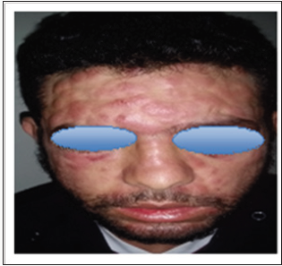
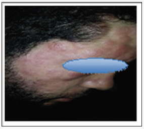
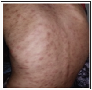
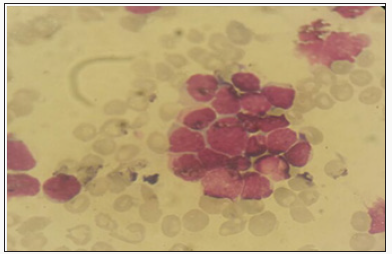
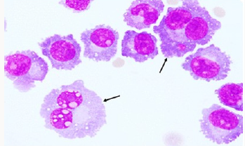
We report the case of 24 years old patient, with no significant pathological history, who had a rash for 10 days in a context of fever and very bad general condition. At admission the patient was febrile, tachycardic and dyspneic. Physical examination revealed erythemato-purplished papulo-nodules on the face, trunk, limbs and a gingival hyperplasia. The oral state was deplorable. Bilateral cervical lymphadenopathy was also found without the patosplenomegaly (Figures 1-3). The biological assessment showed a CRP of 150 and a pancytopenia with a Hbat 7.5g / dl, normal VGM and CCMH, a deep thrombocytopeniaat 85000 / l, leukocytesat 1500 / l. The blood smear showed 35% of circulating blasts and 22% of erythroblasts (Figure 4).
Figure 1: Clinical Manifestations of AML.
Figure 2: Clinical Manifestations of AML.
Figure 3: Clinical Manifestations of AML.
Figure 4: Blood smear showing 35% of circulating blasts.
The medullo gram showed a hyper-cellular marrow with a rate of myeloblasts greater than 45% compared to all non-erythroblastic elements and erythroblastic hyperplasia estimated at more than 65%; with signs of dyserythropoiesis suggestive of the diagnosis of erythroleukemia (Figure 5). Blood immune phenol typing was positive for CD13, CD33, MPO and Glycophotin A. The evolution was unfavourable; the patient died due to massive alveolarhemorrhage.
Figure 5: Hyper cellular marrow infiltrated by a blastic contingent estimated at 45%.

Discussion

Acute erythroblastic leukemia is characterized by the proliferation of a pre dominantery throcyte population on other lineages. There are two types: Erythroleukemia: defined by the presence in the bone marrow of more than 50% of the erythroid precursors of all the medullary cells, and more than 20% of myeloblasts of the whole non-erythrocytemedullary cells - Pure erythroid leukemia: it presents a neoplastic proliferation made of more than 80% of erythrocyte cells without obvious presence of the myeloblastic contingent [1]. It is usually manifested by signs of bone marrow failure and cytopenia [2,3], skin involvement remains rare, varied and disorienting the diagnosis; they are found mainly in Acute myelovlastic leukemia [4,5]. Cutaneous manifestations during leukemia are infrequent and varied. They designate all the cutaneouslesions related to the haematological malignancy directly or indirectly following their treatment; we essentially distinguish.
The specific dermatological lesions which can reveal hematological diseases [4], are mainly represented by leukaemides (leukaemia cutis), which are red brown to purple dermal papules, plaques or nodules. Granulocyticsarcom as an extra-medullary tumour masses, ulcerated plaques and gingival hypertrophy [5]. The infectious dermatoses secondary to the biological disturbances accompanying the malignan themopathy and their treatments. The occurrence of specific cutaneouslesions in leukemia is synonymous with a major aggravation of the prognosis (with for example a survival twice as short if there is a specific cutaneous involvement); this seriousness make some authors propose different treatments with a medium-long stay hospitalization[6-8].
In our case, acute myelonlastic leukemia 6 (AML 6 ) was revealed by diffuse leukemias resulting from the infiltration and proliferation of malignantha ematological cells (blasts) in the skin and by gingival hyperplasia secondary to mucosal infiltration [5]. The clinical presentation of acute leukemia including AML6 in the form of ulcer ativenecroticgingivitis in the foreground, is a rare form to be remembered, mentioned in all courses of medicine and dentistry, stipulating that Gingival involvement is a classic feature of leukemia [6] The frequent association of skin cancers with haematological malignancies is also highlighted in several publications [5].

Conclusion

The cutaneous localizations are among the rarest extreme dullary lesions of acute myeloid leukaemia’s (AML) not exceeding 1%. They are generally considered as factors of worse prognosis. Their cytogenetic or mutational specificities remain un established to date.


For More Lupine Publishers Open Access Journals Please visit our Website:

For more Open Access Journal of Oncology and Medicine Please Click Here: https://lupinepublishers.com/cancer-journal/

 
Follow on Twitter   :  https://twitter.com/lupine_online


Friday, September 13, 2019

Lupine Publishers | Central Nervous System Manifestation of Multiple Myeloma: A Case Report

Lupine Publishers | Open Access Journal of Oncology and Medicine

Abstract

Multiple myeloma accounts for <1% of all malignancies and Central nervous system (CNS) manifestation of multiple myeloma (MM) is rare. Multiple myelomais characterized by malignant transformation of plasma cells that produces immunoglobulin chains. The accumulation of plasma cells in bone marrow results in clinical features like anaemia, osteolytic lesions, hypocalcaemia, renal failure and immunodeficiency [1,2]. The involvement of central nervous system is very uncommon in multiple myeloma and it may present early or late during the course of disease. The neurological symptoms can be diffuse headache, persistent vomiting, vertigo, weakness in limbs, urinary incontinence etc. Symptoms can be attributed to spinal cord or nerve root compression, peripheral neuropathies, and uremia [3,4]. Likely risk factors of CNS manifestation are unfavourable cytogenetics, high tumour load, low marrow involvement. In this report we describe a patient with CNS involvement after receiving systemic chemotherapy and symptomatic treatment.
Keywords: Central nervous system, Multiple myeloma

Case Report

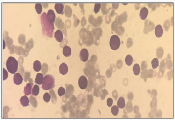
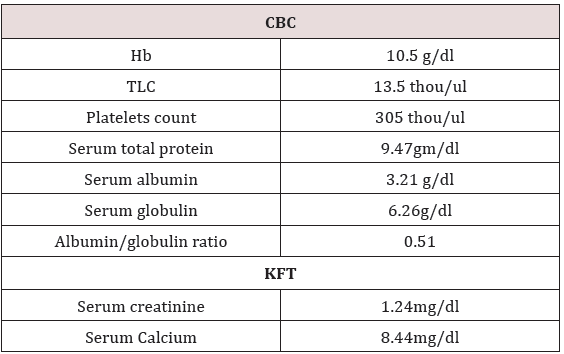
A 52 years pleasant lady presented with complaint of pain in left leg with lower backache and tingling sensations for 10 days. Subsequently she developed pain in right leg followed by urine incontinence. MRI Lumbosacral Spine was done which reported ill defined lesions at L3-L4 vertebra (with associated periosseous fluid collections) and multiple focal lesions in the L4 vertebra, bilateral sacral ala and both iliac bones along with indentations upon the thecal sac by spondyliticridges, secondary spinal canal stenosis and neural foraminal narrowing at multiple levels and left exiting nerve root compression at L3-L4, L5-S1 levels (Table 1). She underwent L1-L5 stabilization, L3 decompression with laminectomy, biopsy, posterolateral fusion with local autograft. Histopathology reported plasma cell dyscrasia at L3-L4 level. Immunohistochemistry reported tumor cells are focally positive for CD-138 and kappa light chain and negative for lambda light chain. On further evaluation bone marrow biopsy reported plasma cells approximately 50% of total nucleated cells suggestive of plasma cell myeloma. Bone marrow aspiration shows plasmacytosis approximately 24% of total nucleated cells which confirmed the diagnosis. She received 15 cycles of Bortezomib and Thalidomide.
Table 1:
Lupinepublishers-openaccess-cancer-Oncology
Table 2:
Lupinepublishers-openaccess-cancer-Oncology
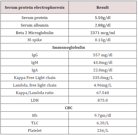
In view of persistent pain in legs MRI dorsal spine was done which reported well defined altered intensity expansile lesion in L3 vertebra extending into epidural space. She received palliative external beam radiotherapy to L3 vertebra 30Gy/10 fractions. She remained asymptomatic for a month. Then she presented with complaint of lower limbs weakness and nausea, loss of appetite, constipation, weight loss (Table 2). USG whole abdomen was done which reported multiple hypoechoic masses in epigastric region. Whole body PET CT reported metabolically active soft mass lesion in gastro hepatic region measuring 9.7x5.5 cm SUV max 20.3, recto uterine pouch measuring 9.2 x 7.8cm SUV 20.9, left iliac fossa measuring 7.2x4.0cm- ?mitotic, metabolically active mesenteric nodes measuring upto 2.2cm-? mitotic and metabolically active lytic lesion involving L3 vertebra with paravertebral soft tissue component infiltrating left psoas muscles.
Table 3:
Lupinepublishers-openaccess-cancer-Oncology
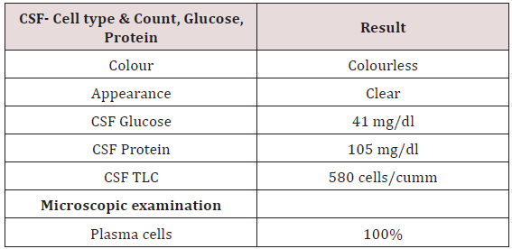
Table 4:
Lupinepublishers-openaccess-cancer-Oncology
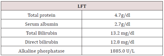
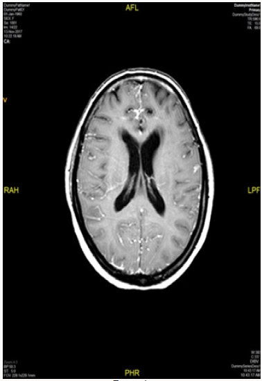
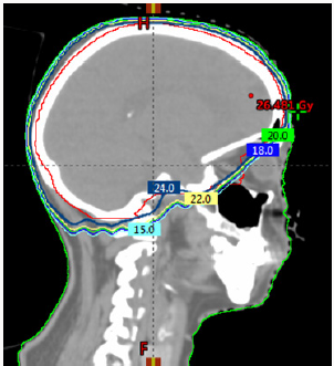
To confirm the diagnosis CT guided core needle biopsy from supraumbilical mass was done which was compatible with multiple myeloma. She then received 1st cycle of Bortezomib, cyclophosphamide and dexamethasone. After few days, she developed headache, giddiness and syncopal attacks. On evaluation CEMRI brain reported diffuse leptomeningeal enhancement involving both cerebral as well as cerebellar hemispheres. Mild enhancement of basal cistern with associated marginal dilation of ventricular system (Figure 1). In view of above mentioned complaints, cerebrospinal fluid study was done (Table 3) (Figure 2). CEMRI spine was done which reported no abnormal enhancement in vertebra, cord or thecal sac (Figures 3 & 4). She received 7 fractions (14 Gy) out of 12 planned fractions (24Gy) of whole brain radiotherapy and two intra thecal injection of methotrexate. MRI upper abdomen with MRCP reported large mass replacing entire head and body region of pancreas. Mass encases and compresses CBD with mild bilobar intra hepatic biliary radical dilation. There is also encasement of second part of duodenum with possible infiltrartion of adjacent hepatic parenchyma as well. Features are suggestive of aggressive pancreatic malignanacy, possibility of pancreatic lymphoma. Thickening and edema of gall bladder wall was seen which was suggestive of concomitant cholecystitis. She was advised for ERCP with SEMS placement which was refused by attendants (Figure 5). In view of her deranged liver function test, both radiotherapy and methotrexate were discontinued and she was maintained on supportive care (Table 4).
Figure 1:
Lupinepublishers-openaccess-cancer-Oncology
Figure 2:
Lupinepublishers-openaccess-cancer-Oncology
Figure 3:
Lupinepublishers-openaccess-cancer-Oncology
Figure 4:
Lupinepublishers-openaccess-cancer-Oncology
Figure 5:
Lupinepublishers-openaccess-cancer-Oncology

Discussion

Central nervous system is a rare location of involvement in MM, however it should be considered if a patient with MM presents with neurologic symptoms. Patients with MM often have neurological complications, either due to metabolic disorders such as hypocalcaemia, uraemia and hyper viscosity or due to peripheral neuropathy, spinal cord compression and cranial nerve infiltration [1,2]. The most common cause is spinal cord compression and cranial nerve infiltration [3,4]. The patient may present as a known case of MM with CNS features or as a new case with CNS symptoms. The clinical presentation depends on degree and site of infiltration. The symptoms and signs are headache, memory loss, behavioural changes, convulsions, nausea, vomiting, vertigo, urinary incontinence, backache, and limb weakness [3,4]. These symptoms can be attributed to spinal cord or nerve root compression, raised intracranial tension, meningeal inflammation [5]. In our case she presented with the complaint of headache, giddiness and syncopal attacks. This can be explained by CEMRI brain imaging which reported diffuse leptomeningeal enhancement involving both cerebral as well as cerebellar hemispheres. CSF study was done to reported to be negative. She received 7 fractions out of 12 fractions planned for whole brain radiotherapy and two injections of intrathecal Methotrexate injections [6]. In view of deranged LFT, radiotherapy and IT chemotherapy was stopped and was managed conservatively. She passed away after 2 weeks due to sepsis with LRTI with MM as underlying cause.

Conclusion

In conclusion this patient had an aggressive disease in view of abdominal deposits and unresponsiveness to multiple lines of systemic disease. CNS involvement in cases of MM portends extremely poor prognosis and median overall survival of <6 months. While in this case there was CSF infiltration which would portend even poorer prognosis but she succumbed to sepsis due to Lower respiratory tract infection. CNS disease in MM can be effectively palliated especially because of relative radio sensitivity of MM due to which early responses are seen at lesser doses and an early diagnosis can improve quality of life effectively.

For more Lupine Publishers Open Access Journals Please visit our website: http://lupinepublishers.us/ For more Open Access Journal of Oncology and Medicine Please Click Here: https://lupinepublishers.com/Cancer-journal/
To Know More About Open Access Publishers Please Click on Lupine Publishers


Wednesday, August 28, 2019

Lupine publishers | Head Cancer and Metastasic Neck. What Have We Advanced in the Last Years?

Lupine Publishers | Open Access Journal of Oncology and Medicine


Abstract


In 2012, 5.210 new cases of head and neck tumors were estimated in the USA, with an increasing incidence due to tobacco and alcohol habits in the population. A large percentage of the cases debut as a locally advanced disease, so control of the disease is key and we look for the best therapeutic strategy to achieve good survival rates while maintaining quality of life. We present the case of a 60-year-old patient in which our objectives to be presented are the assessment of comorbidity, toxicity and survival.

Clinical Case

A 60-year-old man without medical illnesses to be highlighted. He came to the emergency room in July 2016 due to injury at the cervical level of 1 month of evolution, with progressive growth and breathness, with also difficulty for eating. Also asthenia, anorexia and loss of weight not quantified in the last month.
Physical Examination: Weight 42 kg Head and Neck: mass of hard consistency of approximately 10 cm in diameter in the cervical left region that seems to deflect trachea. Pulmonary auscultation: generalized hypoventilation with some expiratory wheeze.

Additional Tests

a. Fibroscopy (August 2016) ulcerated lesion from right vallecula to mouth of Killian. Paresis of both vocal cords.
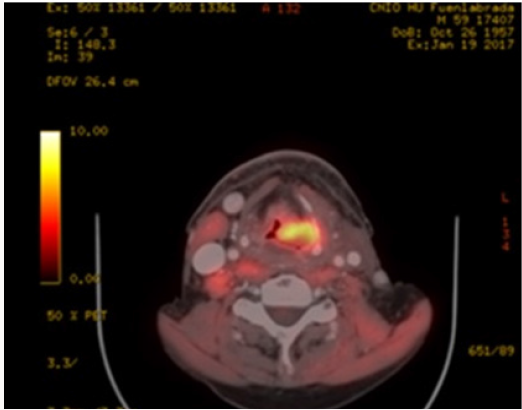
b. PET-CT (August 2016): Extensive tumor in pharynxlarynx- esophagus (> 6 cm) Lymph nodes in left IB-II spaces (> 6 cm). If confirmed, carcinoma would correspond to T3 N3 (stage IVB) (Figure 1).
c. Laryngeal Biopsy: Moderately differentiated and keratinizing squamous cell carcinoma.
Figure 1: PET-CT August 2016: Pathological adenopathies> 6 cm in left IB-II spaces
Lupinepublishers-openaccess-cancer-Oncology
So, confirmed Squamous cell carcinoma stage cT4N3 Mx.The case is presented in the Tumor Committee, deciding treatment with Chemoradiotherapy and 1 cycle of Docetaxel+ Cisplatin (60% dose reduction because of frailty/ malnutrition) previous to induction due to the large tumor volume and waiting for start radiotherapy. Tracheotomy is performed prior to starting because of the risk of airway obstruction and also gastrostomy is placed for nutritional support.
On 31th August 2016 the patient starts on RT concomitantly to Cisplain ( receiving 2 cycles on 12.09.2016 and 10.10.2016 and 70 Gy) During the treatment, he achieves a good general condition until January 2017, when he goes to Otolaryngology Clinics referring dysphagia again although he maintains weight in 51 kg. PET-CT is performed: disease progression with soft tissue increase in the pharyngoesophageal junction despite the good response of cervical adenopathies and the partial response of the primary tumor. New bilateral subpleural pulmonary nodules suggestive of metastasis. Figure 2 Due to progression, he restarts treatment with chemotherapy (palliative intention) with ERBITAX scheme (Paclitaxel 80 mg / m2 weekly (3 / 4s) + Cetuximab 400 mg / m2 followed by 250 mg / m2) with good tolerance,only highlighting secondary rash to cetuximab (predictive factor). After 3 cycles he presents significant partial response (Figure 3) and continues until 6 cycles. After 10 cycles it is considered whether to stop paclitaxel and follow on with cetuximab, but given the good tolerance they remain both of them. However, in January 2018, he presented a new pulmonary progression, so we decided to start a new strategy with inmunotherapy (Nivolumab), receiving 2 cycles to date.
Figure 2: PET-TC January 2017: locoregional tumor progression.
Lupinepublishers-openaccess-cancer-Oncology
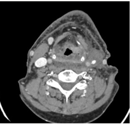
Figure 3: TC March 2017: Partial response of left adenopathy
Lupinepublishers-openaccess-cancer-Oncology

Discussion

a. The concept of fragility is evaluated incorrectly through the Performance Status (PS). The ACE scale 27 assesses comorbidities and compares survival to having an advanced stage, so that is a fact to take into account more than the ECOG at the time of choosing the treatment.
b. When a patient progress to chemoradiotherapy, we have two good “palliative chemo” options: phase III EXTREME study(5-FU + Cisplatin + Cetuximab) or phase II of Hitt (Paclitaxel-Cetuximab) with fewer side effects, which is an alternative to cisplatin, achieving good response rates (20.43%) [1].
c. After 6 cycles of Paclitaxel + Cetuximab can be considered to keep Cetuximab as monotherapy, continue both or suspension until progression.
d. If pulmonary metastatic disease is controlled for> 1 year, we can consider surgical intervention.
e. Support treatment improves tolerance to chemotherapy. The indication of prophylactic enteral nutrition is a controversial issue, so we have to individualize.
f. We need predictive factors for each tumor type that we can know about before hand the prognosis and guide the treatment according to it. We must raise multidisciplinary strategies with the aim of achieving the best treatment sequence to improve survival in a population with few therapeutic options [2]

For more Lupine Publishers Open Access Journals Please visit our website:
http://lupinepublishers.us/
For more Open Access Journal of Oncology and Medicine Please Click Here:
https://lupinepublishers.com/Cancer-journal/

To Know More About Open Access Publishers Please Click on Lupine Publishers

Wednesday, July 17, 2019

Oncology and Medicine Journals - Lupine Publishers

What is beyond the Nivolumab Monotherapy approval for advanced Hepatocellular Carcinoma? By Luis Mendoza In Open Access Journal of Oncology and Medicine (OAJOM)- Lupine Publishers


With an estimated 500,000 new cases per year, hepatocellular carcinoma (HCC) represents the third leading cause of cancer death worldwide. The incidence is rising in the west, largely due to an increasing incidence of hepatitis C virus infection [1]. The majority of HCC patients are diagnosed with disease too advanced for curative treatment. Only liver resection and liver transplantation are considered curative, with poor efficiency of other modalities such as radiofrequency ablation (RFA) and transarterial chemoembolization (TACE), although this may provide a modest prolongation in survival; however, the relapse in the majority of these patients is inevitable [2]. An array of translational research and pilot clinical trials have revealed that adoptive immunotherapy’s are safe by patients with HCC, but they lack efficacy [3]. Now, we are in the new era of immunotherapy’s such as immune checkpoint inhibitors and CAR-T strategies, which would bring benefit to the HCC patients.On September 22, 2017, the Food and Drug Administration granted accelerated approval to nivolumab (OPDIVO, Bristol- Myers Squibb Co.) for the treatment of HCC in patients who have been previously treated with sorafenib. The approval was based on a 154-patient subgroup of CHECKMATE-040 (NCT 01658878), a multicenter, open-label trial conducted in patients with HCC and Child-Pugh. A cirrhosis who progressed on or were intolerant to sorafenib. Patients received nivolumab 3 mg/kg by intravenous infusion every two weeks. The confirmed overall response rate, as assessed by blinded independent central review using RECIST 1.1, was 14.3% (95% CI: 9.2, 20.8), with three complete responses and 19 partial responses. The response duration ranged from 3.2 to 38.2+ months; 91% of responders had responses lasting six months or longer and 55% had responses lasting 12 months or longer. Adverse reactions occurring in patients with HCC in CHECKMATE-040 were similar to those previously reported in product labelling, with the exception of a higher incidence of elevations in transaminases and bilirubin levels [4].

For More Information
https://lupinepublishers.com/cancer-journal/fulltext/What-is-beyond-the-nivolumab-monotherapy-approval-for-advanced-hepatocellular-carcinoma.ID.000109.php

For more Lupine Publishers Open Access Journals Please visit our website: https://lupinepublishersgroup.com/

For more Oncology and Medicine Journals  articles Please Click
Here: https://lupinepublishers.com/cancer-journal/

Wednesday, July 10, 2019

Oncology and Medicine Journals - Lupine Publishers


As oncologists learn to target the immune response to “self and non-self,” a delicate therapy balance will eventually be achieved with predictable outcomes, benefits, and toxicity in the fightThe study of how the immune system recognizes friend and foe, or as the immunologist Sir Macfarlane Burnet phrased it, “distinguishes between self and non-self,” has driven important discoveries that are transforming our ability to treat cancer. Over the last few clinicians have unraveled the interactions (both innate and adaptive immunity) that lead to the eradication of viruses, bacteria, parasites, and now, cancer. Notable cellular players include T cells, B cells, natural killer (NK) cells, neutrophils, eosinophils, basophils, dendritic cells, and macrophages, along with a host of secreted mediators - antibodies, complement, cytokines, and chemokines - each of which fulfills particular immunologic functions. Processes, autoimmune disease can be a consequence. These diseases also occur if shared. When the immune system fails to regulate these antigens are recognized by the immune system in cells; one example is Lambert-Eaton syndrome. Monoclonal antibodies that target tumour reactive T cells (eg, nivolumab and pembrolizumab) can also cause autoimmune disease; other examples include graft-vs-host disease (GVHD) in allogeneic bone marrow transplant recipients and cytokine release syndrome (CRS), which is associated with adoptive T cell therap.

For More information
https://lupinepublishers.com/cancer-journal/fulltext/cancer-and-advantages-of-immunosuppression.ID.000108.php

For more Lupine Publishers Open Access Journals Please visit our website: https://lupinepublishersgroup.com/
 
For more Oncology and Medicine Journals  articles Please Click
Here: https://lupinepublishers.com/cancer-journal/

Wednesday, June 19, 2019

Oncology research journals - Lupine Publishers

Targeting the Immune Checkpoint in Cancer: Is This a Viable Treatment Option for AML? by Steven J Coles in Open Access Journal of Oncology and Medicine (OAJOM) - Lupine Publishers

The immune suppressive mechanisms displayed by malignant cells are considered a central process in the pathogenesis of cancer. Research in this area has gained significant momentu mover the past 20 years, with several immune checkpoints identified, including; CTLA-4, CD200/CD200R, Tim-3/Galectin-9 and PD-L1/PD-1 (Figure 1). Whilst characterising the molecular basis of leukaemia for risk stratification remains at the forefront of AML research; this must now extend to understating how the seimmune checkpoint path ways fit into the equation. A good example of why this is important is to consider CD200expression level in AML, which is a negative prognostic indicator [1]. CD200 is an immunosuppressive lig and, that when engaged with its receptor CD200R, has the capacity to attenuate T-cell and NK-cell anti-tumour activity in AML. Interestingly, most cases of CBF AML express high levels of CD200, yet CBF AML performs relatively well clinically. This paradox suggests there is a complex interplay between AML molecular heterogeneity and immune surveillance. Given the recent development and FDA approval of several immune checkpoint therapies, a full understanding of these processes and integration with standard molecular risk stratification is warranted.

For More information

For more Lupine Publishers Open Access Journals Please visit our website
For more Oncology research journals  articles Please Click
Here: https://lupinepublishers.com/cancer-journal/

Lupine Publishers | Open Access Journal of Oncology and Medicine (OAJOM)

      Thanksgiving   is a national   holiday   celebrated on various dates in the United States, Canada, Grenada, Saint Lucia, and Liberia. ...