Showing posts with label Lupine PubliLupine Publishers open access journals. Show all posts

Tuesday, May 26, 2020

Lupine Publishers | A Case Report and Review of Thymic Carcinoma with Adenoid Cystic Carcinoma like Features

Lupine Publishers | Open Access Journal of Oncology and Medicine


Introduction

Adenoid cystic carcinoma (ACC) is also called cylindroma, which is considered as a kind of low-grade malignant tumor and often occurs in head and neck salivary gland tissue. Most of them happened in submandibular gland and minor salivary glands while rarely happened in thymus. There are only 7 cases had already reported before all around the world. Our case is diagnosed as thymic carcinoma with adenoid cystic carcinomalike features by pathology after surgery; it is different with other cases because he had accepted a surgery six years ago because of the mediastinal mass and pleural effusion. Because of the symptom of chest tightness, he came to see doctor again and finally find there was a mediastinal mass and accepted the treatment of surgery. The medical history of our case maybe provides some inspiration of the pathogenic of thymic carcinoma with adenoid cystic carcinoma like features.

Information and history

A 60 years old male came to our hospital because of chest tightness in 2009/10. The enhanced chest CT showed “A large mass measuring 9.2*10.1cm² in the right side of the mediastinal which the average CT value is 9 Hu. The border with the superior vena cava and the right atrial and mediastinum is less clear. The right side of the chest is hydropneumothorax, Hydropericardium. The ultrasonic showed there is a mass of mixed echo in the right chest which had strong echo light and liquid visible mass dark space inside it. The test of pleural effusion by pleural puncture showed that was hematodes exudates without cancer cells. He accepted a surgery to resection of the mass by posterolateral thoracotomy, the pathology showed “Mostly considered as hematoma with organization according to clinical”. After the surgery ,he recovered and discharged from our hospital.
It is said by himself that there was found a mediastinal mass in his chest by CT in the year of 2010(the imaging data has been lost), and he taking chinese traditional medicein(herbal medicine) for tow month. After that he reexamination a chest CT showed the mediastinal mass was disappeared so he didn’t have the follow-up according to the doctor’s advice. 2014/10/13, he had a chest CT examination showed there is a 15*13cm² cystic with solid abnormal density shadow in the right front mediastium; Mediastinal lymph node is enlarged; Density of soft tissue in chest wall which is considered as metastatic. He reexamined a chest CT in 2015/1/4 but did not show significant change. Finally, he came to hospital again because of the intermittent chest pain. Complete the checks after hospitalization, lung function show” FEV1 : 3.94L, FEV1/FVC : 79%”, the enhanced chest CT showed “there is a 15.2*11.6cm² cystic with solid abnormal density shadow in the right front mediastium, the border is smooth and the density inside is uniform, the CT value is 18Hu; Right pulmonary atelectasis, the chest wall and diagram pars maybe have already metastasis tumor”.
a. Anamnesis: The patient with high blood pressure for more than 20 years, he use reserpin and Chinese traditional medicine to ctrl his high blood press by himself, but the effect is poor. His blood pressure was stabled for able 108/90mmHg for all years. Smoking 20 years and quit it for 23 years. We asked his medical history repeatedly, he denied the history of trauma and hemorrhagic disease.
b. Preoperative diagnosis: anterior mediastinum space occupying lesion, malignant tumor is the most possible. The chest wall maybe already metastasized. We suggested him to accept PET/CT to make the tumor’s metabolic strength clear and conform the primary focal; Or accept thoracentesis by fine needle to definite the pathological diagnosis. The patient and his family refused it and screamed for an operation.
So the surgery was planned. The patient was placed in the supine position under general anesthesia. Via median sternotomy. We saw the tumor was located in right anterior mediastinum, 12*12*10cm³, it was dense adhesion with mediastinal pleura, pericardium, diaphragm and right lung. The tumor is invading and partly wrapped around superior vena cava. Cut out part of the tumor tissue and sent to the examination of fast frozen section and the result was benign tumor. Then we completed removal of the tumor and cut one right rib and the tumor on the chest wall, the fast frozen section also report “benign tumor”. The tumor was solid and multicystic hemorrhagic with many sediment sample material. The diagnosis of the frozen section considered that most possible is encapsulated empyema. So we use polyninylpyrrolidone and normal saline to rinse the chest repeatedly and the closed thoracic conventionally.
The result of postoperative pathological: thymic carcinoma with adenoid cystic carcinomalike features with chest wall invasion, the tissue of cystiform is hyperplasia of fibrous tissue and stale hemorrhage. The result of immunohistochemical : CK(+), EMA(-), CK(L)(+), E-Ca(++), ER(+), PR(-), Ki67(10%), CK8/18(+), CD56 Focal(+), NSE(-), Syn(-), CgA(-).After the surgery, we checked the patient’s oral cavity, head and neck region but not found and obvious abnormal. 6 month pasted and we have not found any indication of tumor recurrence. He is still in follow up now.

Discussion


Adenoid cystic carcinoma is salivary gland tumor, but can still be reported in breast [1], lung [2], esophageal [3], postate [4]. Thymic carcinoma with adenoid cystic carcinoma like features is very rare. Only 7 cases have already be reported all around the world before while all cases is not in China.
Thymic carcinomas are defined as thymic epithelial neoplasmas, which are classified into 10 different histological types according to the 2015 update of the WHO classification, i.e.[5]
a. squamous-cell carcinoma
b. basal cell carcinoma
c. mucoepidermoid carcinoma
d. lymphoepithelioma-like carcinoma
e. sarcomatoid carcimoa
f. clear cell carcinoma
g. adenocarcinoma
h. NUT carcinoma
i. unclassified carcinoma
j. other rare thymic carcinoma.
Within the group of adenocarcinoma, four histologic subtypes are known: papillary adenocarcinoma; thymic carcinoma with adenoid cystic carcinomalikefeatures; mucinous adenocarcinoma; adenocarcinoma, non specifically. Compared with the 2004 update of the WHO classification, the name of adenoid cystic carcinoma is changed to thymic carcinoma with adenoid cystic carcinomalike features. It lack the real features of adenoid cystic carcinoma in the character of immunohistochemical because it is generate from the thymus (Table 1) [6-8].
Table 1: The character of the cases of thymic carcinoma with adenoid cystic carcinomalike features which are reported so far.
Lupinepublishers-openaccess-cancer-Oncology

The Character of Clinical

All of the clinical character of the cases which have already been reported is summarized above (chart-1). Thymic carcinoma with adenoid cystic carcinoma like features is very rare, most of them are occurred in elderly and few distant metastases. But it can show the character of aggressive growth and directly invasive chest wall; pericardium and other adjacent tissue. The initial symptoms of the patients are fever; cough; dyspnea; chest pain; the sense of suppression in the chest and the loss of weight [9]. thymic carcinoma with adenoid cystic carcinoma like features is generated in the tissue of thymus which located in the mediastinum, and the size of the most tumor is large. The severity of the clinical symptoms is uncorrelated with the size of the tumor.
Although the size of the tumor is large, the symptoms of patients is not severely even cannot let them go to see doctor. The clinical symptoms is formed because of the oppress by the tumor, there is no obviously symptoms in early stage, with the growth of tumor , the patient will appear the atypical symptoms such as cough; fever; dyspnea; chest pain and so on. Because thymic carcinoma with adenoid cystic carcinomalike features is a low grade malignancy, all cases had the features of gradual onset and grow progress. So only one case that had already metastases to rib was reported that is different whit the character of adenoid cystic carcinoma in salivary. According to the Masaoka stage of thymus tumor, our case is in stage β.

The Character of Pathology

The macroscopic view of the tumor: Most of them are consist of huge gritty and firm cystic solid mass with smooth border. Some of them have complete capsule. The cross-section showed multicystic, necrotic tissue mixed with fibrotic, yellow tissue nodules. Some of them can be observed the papillary hyperplasia and plenty of hemorrhagic effusion.
a. The microscopic view of the tumor: The character of our case is similar as salivary adenoid cystic carcinoma. The cystic and cribriform texture neoplastic nest is consist of basoloid cell carcinoma ,and bloody fluid or granular basophilic myxoid stroma full of the cyst. The microscopic view of the thymic carcinoma with adenoid cystic carcinomalike features can be classified as three types. If the tumor cell is undifferentiated, it will show the solid structure with irregular funicular or dense array by basaloid cells(solid type). If the tumor cell is differentiation to the glandular epithelium, it will show the gland tubular structure consist of the inner layer of columnar epithelium and the outside of the myoepithelial cells with homogeneous eosinophilic mucin full of the cavity of the tubule(tubular type). If the tumor cell is differentiation to the myoepithelial cell, it will show the cribriform structure with homogeneous basophilic cell matrix(cribriform type)[10]. The classify of thymic carcinoma with ACC like features is closely connected with the prognosis, so we infer that the prognosis of solid type is the worst while the prognosis of tubular type and the cribriform type is more better.
b. Immuno histochemistry: CK and CKL all showed positive in our case, they were marked at the epithelium which consist of cribriform structure. E-Ca(++) prompted the tumor has a certain invasive in accordance with the invasion of chest wall. Ki67(10%) prompted the active proliferation. CD56(focal +) is the new character that has not be reported before ER(+) prompted this case may be sensitive to endocrine therapy, but there isn’t any evidence of evidence-based medicine support it so the clinical significance needs to be confirmed.
Summarize other documents , we can get the following conclusion:
i. These marks is contribute to the diagnosis of thymic carcinoma with adenoid cystic carcinomalike features if they are positive: CollagenIV, laminin, P63, CK34betaE12, Ki67(1- 10%)
ii. These marks is contribute to the diagnosis of thymic carcinoma with adenoid cystic carcinomalike features if they are negative: Syn, NSE, CD117, CgA.
The molecular marks research of this disease has not yet been carried out. There are reports that suggested MYB-NFIB fusion gene, NF-κB, MMP-2, survivin is related to adenoid cystic carcinoma [11], and it is helpful to the molecular marks research of thymic carcinoma with adenoid cystic carcinomalike features.

The Differential Diagnosis

PET/CT has great significance to this disease in differential diagnosis, it can estimate whether the mass is primary tumor or metastases. The report suggested the tumor of this disease show high value of SUV max in PET/CT while its metastases can also show unusually high value of SUV max [9]. The retrospective analysis show that have a examination of PET /CT before surgery is correct and meaningful. In the primary thymic carcinoma, this disease should be distinguished with basaloma. In morphology, most of the tumor cell of basaloma is cubic cells with small and deep dyeing nucleus which array form lobulated or funicular [12,13]. The results of Immuno histochemistry can confirm that our case can be diagnosed as thymic carcinoma with adenoid cystic carcinoma like features.

The treatment and prognosis

Surgery is given priority to this disease at present and radiation therapy can according to the situation. The effect of chemotherapy for this disease is poor and if take chemotherapy as a routine treatment is still controversial. Some professional insist of that adenoid cyst carcinoma cannot get enough benefit from chemotherapy, so we should treat chemotherapy as the last treatment [14]. We believe that A case report and review of thymic carcinoma with adenoid cystic carcinoma like features is a low level malignant tumor so the it should have better prognosis after standardized treatment. But we still should be carefully evaluated before surgery and if we suspect the diagnosis as thymic tumor with ACC like carcinoma or huge mediastinal mass, PET/CT will be suggested to confirm the quality of the mass and whether there is metastases or not.
Locally metastasis and the chest wall invasion should not be the absolute contraindications of surgery because as a low level malignant tumor, if we resection the tumor and metastases completely and take some radiation therapy appropriately, nice quality of life will get. In our case a recurrence is appear after the surgery in 2009, but he said itself that the mediastinal mass was once disappeared after the treatment of Chinese traditional medicine. So we guess that Chinese traditional medicine may effective for this disease.

The discussion of pathogenesis

A case report and review of thymic carcinoma with adenoid cystic Carcinoma like features is a kind of thymic carcinoma, it is generated from thymic epithelial cells. If the tumor cell is differentiation to the myoepithelial cell, it will show the cribriform structure with homogeneous basophilic cell matrix. The medical history of our case is so specially that he found the mediastinal mass six years ago and experienced the surgery. The pathological report at the time of 2009 showed “hematoma with organization”. Organization means the absorption process by new granulation tissue if necrotic tissue, thrombus, .com or foreign body cannot be dissolved or absorbed or separated or discharged. The granulation tissue contains abundant myocyte and some of them will differentiate to myofibroblast after the stimulate of cytokines.
And these cytokines that can induce differentiation may induce the differentiate thymic epithelial cell to myoepithelium and finally generate to the type of cribriform of thymic carcinoma with adenoid cystic carcinomalike features is a kind of thymic carcinoma. This maybe one of the triggers of this disease although still need to establish by experiments. According to the inference above, we consider coagulate hemothorax and mediastinal hematoram may result thymic carcinoma with adenoid cystic carcinoma like features. In order to avoid the remained thymus tissue be stimulated to differentiate to thymic carcinoma, in the surgery of mediastinal hematoma, thymectomy completely is necessary and fat tissue of mediastinal should be resection completely too.

Read More Lupine Publishers Oncology and medicine Articles : 

Read More Lupine Publishers blogger Articles : 
To Know more Open Access Publishers Click on Lupine Publisher:
Follow on Linkedin : https://www.linkedin.com/company/lupinepublishers
Follow on Twitter   :  https://twitter.com/lupine_online 


Wednesday, May 13, 2020

Lupine Publishers | Promising Role of Fractional Calculus in Biomedicine and Biophysics

Lupine Publishers | Open Access Journal of Oncology and Medicine


Introduction
The study of complex systems and investigation of their structural and dynamical properties have attracted considerable interests among scientists in general and physicists, biologists and medical researchers in particular. Complex systems can be found almost everywhere however the highest level of complexities is related to living and biological organisms and systems. Due to the lack of a reliable and effective tool to investigate such systems, we have not reached to the complete understanding and comprehensive pictures of the phenomena and processes which occur in these systems. Of course a comprehensive knowledge of biological and biomedical complex phenomena will be achieved when we employ simultaneously different field of science and engineering including: biology, chemistry, physics, mathematics, mechanical engineering and so on.
Fortunately in recent year's powerful tool of fractional calculus has been proposed for study of complex and nonlinear phenomena. It is in fact very useful tool for describing the behavior of nonlinear systems which are characterized by: special kind of non-locality, long-term memory and fractal properties. There exist many biological objects and systems with memory, nonlocal effects and nonlinear behaviors and such these non-localities and memory effects in biological objects and systems mean that the next state of the organism or system relies not only on its present state but also upon all of its previous states. As a result, the concept of fractional dynamics and in fact adopting fractional calculus can play an important role in the study of dynamical biological systems. Up to now few number of important issues such as: protein folding phenomena and mechanics of cancer cells (for more details see the references which have investigated physics of protein and physics of cancer in detail) have been investigated using the framework of fractional dynamics [1].
However many other important issues still remain as open issues, such as: modeling of interactions between light (laser) and biological tissue and modeling of intracellular (and intercellular) interactions in the framework of fractional dynamics. As a physicist or biologists and even medical researchers, we always are able to model natural phenomena for instance modeling of tumor growth using systems of differential equations and nowadays it is well know that the fractional-order ones are more comprehensive and also incorporate memory effect and the concept of non-locality in the model.
Mathematically the idea is in fact, to rewrite the ordinary governing differential equations in the fractional form by replacing the standard derivative with a fractional derivative of arbitrary order which is defined in the Caputo sense as follows:
where Γ denotes the Gamma function and , . And its Laplace transform can be given by:

Where, F(s) is the Laplace transform of f (t). Solutions of fractional differential equations generally will be expressed using a generalized special function named as Mittag-Leffler function. This function can be considered as a generalized exponential function and has several different forms. For instance the one-parameter Mittag-Leffler function is defined by the series expansion as:
Where 
C is the set of complex numbers? It is worth mentioning that the exponential function is just a special case of α = l Mittag-Leffler function, for example for the special case of , the Mittag-Leffler function Eq. (3) reduces to the exponential function E
1(z) = ez . This point is very important because of that the natural exponential function has been considered as a fundamental function of natural science and in particular biology up to now, so that many phenomena could be described using it and now scientist are able to think that with such this new framework (i.e. fractional differential equations and their solutions in terms of Mittag-Leffler functions) they can find many new results and information about biological and biomedical phenomena [2,3].
Finally, based on all above mentioned reasons, as a conclusion we should say that we believe that the powerful tool of fractional calculus and in fact the frame work of fractional dynamics can give.com new insights in understanding and modeling of nonlinear complex phenomena in various living cellular structures and their interactions and we invite all biologist and medical researchers to consider this new powerful approach for their future studies.

Read More Lupine Publishers Oncology and medicine Articles : https://lupine-publishers-cancer-journal.blogspot.com/

Read More Lupine Publishers blogger Articles : https://lupinepublishers.blogspot.com

To Know more Open Access Publishers Click on Lupine Publisher

Follow on Twitter   :  https://twitter.com/lupine_online  

Thursday, March 12, 2020

Lupine Publishers | Acute Erythroblastic Leukemia Revealed by Dermatological Manifestations

Lupine Publishers | Open Access Journal of Oncology and Medicine

Abstract

Acute erythroblastic leukemia is characterized by the proliferation of a predominant erythrocyte population on other lineages. Cutaneous manifestations remain rare and misleading, making the diagnosis of difficult to suspect as first-line. Here, we report an unusual and rare case of acute leukemia in a 24 year old male with gingival hypertrophy and dermatological manifestations. This case emphasizes that dentist and dermatologist should be well acquainted with these manifestations of systemic diseases.

Case Report

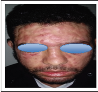
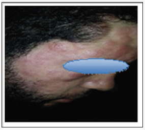
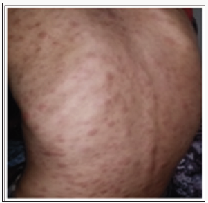
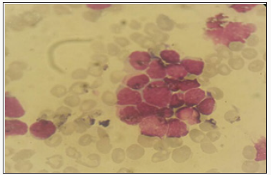
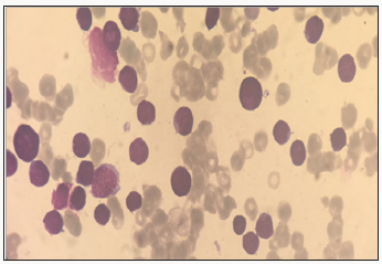
We report the case of 24 years old patient, with no significant pathological history, who had a rash for 10 days in a context of fever and very bad general condition. At admission the patient was febrile, tachycardic and dyspneic. Physical examination revealed erythemato-purplished papulo-nodules on the face, trunk, limbs and a gingival hyperplasia. The oral state was deplorable. Bilateral cervical lymphadenopathy was also found without the patosplenomegaly (Figures 1-3). The biological assessment showed a CRP of 150 and a pancytopenia with a Hbat 7.5g / dl, normal VGM and CCMH, a deep thrombocytopeniaat 85000 / l, leukocytesat 1500 / l. The blood smear showed 35% of circulating blasts and 22% of erythroblasts (Figure 4).
Figure 1: Clinical Manifestations of AML.
Figure 2: Clinical Manifestations of AML.
Figure 3: Clinical Manifestations of AML.
Figure 4: Blood smear showing 35% of circulating blasts.
The medullo gram showed a hyper-cellular marrow with a rate of myeloblasts greater than 45% compared to all non-erythroblastic elements and erythroblastic hyperplasia estimated at more than 65%; with signs of dyserythropoiesis suggestive of the diagnosis of erythroleukemia (Figure 5). Blood immune phenol typing was positive for CD13, CD33, MPO and Glycophotin A. The evolution was unfavourable; the patient died due to massive alveolarhemorrhage.
Figure 5: Hyper cellular marrow infiltrated by a blastic contingent estimated at 45%.

Discussion

Acute erythroblastic leukemia is characterized by the proliferation of a pre dominantery throcyte population on other lineages. There are two types: Erythroleukemia: defined by the presence in the bone marrow of more than 50% of the erythroid precursors of all the medullary cells, and more than 20% of myeloblasts of the whole non-erythrocytemedullary cells - Pure erythroid leukemia: it presents a neoplastic proliferation made of more than 80% of erythrocyte cells without obvious presence of the myeloblastic contingent [1]. It is usually manifested by signs of bone marrow failure and cytopenia [2,3], skin involvement remains rare, varied and disorienting the diagnosis; they are found mainly in Acute myelovlastic leukemia [4,5]. Cutaneous manifestations during leukemia are infrequent and varied. They designate all the cutaneouslesions related to the haematological malignancy directly or indirectly following their treatment; we essentially distinguish.
The specific dermatological lesions which can reveal hematological diseases [4], are mainly represented by leukaemides (leukaemia cutis), which are red brown to purple dermal papules, plaques or nodules. Granulocyticsarcom as an extra-medullary tumour masses, ulcerated plaques and gingival hypertrophy [5]. The infectious dermatoses secondary to the biological disturbances accompanying the malignan themopathy and their treatments. The occurrence of specific cutaneouslesions in leukemia is synonymous with a major aggravation of the prognosis (with for example a survival twice as short if there is a specific cutaneous involvement); this seriousness make some authors propose different treatments with a medium-long stay hospitalization[6-8].
In our case, acute myelonlastic leukemia 6 (AML 6 ) was revealed by diffuse leukemias resulting from the infiltration and proliferation of malignantha ematological cells (blasts) in the skin and by gingival hyperplasia secondary to mucosal infiltration [5]. The clinical presentation of acute leukemia including AML6 in the form of ulcer ativenecroticgingivitis in the foreground, is a rare form to be remembered, mentioned in all courses of medicine and dentistry, stipulating that Gingival involvement is a classic feature of leukemia [6] The frequent association of skin cancers with haematological malignancies is also highlighted in several publications [5].

Conclusion

The cutaneous localizations are among the rarest extreme dullary lesions of acute myeloid leukaemia’s (AML) not exceeding 1%. They are generally considered as factors of worse prognosis. Their cytogenetic or mutational specificities remain un established to date.


For More Lupine Publishers Open Access Journals Please visit our Website:

For more Open Access Journal of Oncology and Medicine Please Click Here: https://lupinepublishers.com/cancer-journal/

 
Follow on Twitter   :  https://twitter.com/lupine_online


Thursday, February 6, 2020

Lupine Publishers | Somatic Mutations in Cancer-Free Individuals: A Liquid Biopsy Connection

Lupine Publishers | Open Access Journal of Oncology and Medicine


Abstract

Somatic mutations have been perceived as the causal event in the origin of the vast majority of cancers. Advanced massively parallel, highthroughput DNA sequencing have enabled the comprehensive characterization of somatic mutations in a large number of tumor samples for precision and personalized therapy. Understanding how these observed genetic alterations give rise to specific cancer phenotypes represents an ultimate goal of cancer genomics. However, somatic mutations are also commonly found in healthy individuals, which interfere with the effectiveness for cancer diagnostics.
Keywords: Somatic mutation; Germline; Cell-free DNA; Liquid biopsy; Next-generation sequencing
Abbreviations: NGS: Next-Generation Sequencing ; cfDNA: Cell-free DNA; MAF: Mutant Allele Frequency

Introduction

Mutations in healthy individuals are not all germline
Over the course of our lifetime, there are many millions of cell divisions in the body. By chance alone, mutations will definitely occur. Indeed, spontaneous somatic mutations constantly occur in individual cells. These background mutations arise either from replication errors or from DNA damage that is repaired incorrectly or left unrepaired, and have been detected in healthy tissues, including blood, skin, liver, colon, and small intestine [1-3]. Deepsequencing studies in normal tissues also surprisingly identified cancer-driving mutations, e.g., in blood, driver mutations can be detected in ~10%of individuals older than 65 years of age and resemble patterns seen in leukemia patients. Individuals carrying these driver mutations have an elevated future risk of blood cancers [4-6], suggesting that these are genuine precancerous clones. Further, a detailed analysis of 31,717 cancer cases and 26,136 cancer-free controls from 13 genome-wide association studies revealed that the majority, if not all, of aberrations that were observed in the cancer-associated cohort were also seen in cancer-free subjects, albeit at lower frequency [7,8].
Somatic mutations in healthy individuals are very prevalent, with an average mutation number of around 2–6 mutations/1 M bases [9,10]. The baseline somatic mutation spectrum in healthy population not only can help fill the gaps for the establishing early cancer diagnosis strategies, but also argues against the idea of using normal cells as germline control to make somatic mutation calls in sequencing tests. Moreover, the same driver mutation could exist in both tumor and normal cells yet with distinct biological effects, we should not simply define the threshold of mutation detection by removing the background mutations found in a healthy population. Taken together, we need to incorporate and carefully calibrate the background somatic mutations in healthy individuals; the fact is they are not all germline mutations.
Somatic driver mutations found in healthy population by liquid biopsy
With the dramatically decreased cost of next-generation sequencing (NGS) in recent years, it is now practical to screen a large number of individuals at ultra-deep sequencing depths to identify cancer-related mutations. Cell-free DNA (cfDNA) in the blood circulation of cancer patients (as liquid biopsy) have emerged as key biomarkers for cancer monitoring and treatment decisionmaking [11]. Both academic research groups and industry players are chasing the pan-cancer screening by a simple blood draw. However, the reliable and accurate application of cfDNA detection requires better understanding of background somatic information in healthy individuals.
We performed ultra-deep target sequencing on 50 cancerassociated genes for plasma cfDNA from a cohort of 129 apparently healthy cancer-free subjects. To increase the confidence of the called mutations, we here defined the mutation as the variant allele frequency greater than 1% and the average depth more than 5,000 xs for demonstration. Our data revealed an age-independent mutation spectrum with average 3.12 somatic mutations per subject (Figure 1). The most frequently mutated genes are TP53 (42%), KIT (6%), KDR (5.5%), PIK3CA (5.5%), EGFR (5%) and PTEN (3.7%). These results highlighted the prevalence of some cancer-associated driver mutations in healthy individuals as background mutations. We also demonstrated the concordance between our results and a recent study for revealing the real somatic mutation in healthy population.
Figure 1: Distribution plots of somatic mutation detected in a cohort of 129 healthy subjects.
The study by Xia et al. [12] examined the background somatic mutations in white blood cells and cfDNA in healthy controls based on sequencing data from 821 non-cancer individuals with the aim of understanding the baseline profile of somatic mutations detected in cfDNA. The data comparison was summarized in Figure 2. Although there are differences in study cohort composition, sample volume, extraction methodology and analytical platform, the end results are remarkably similar, i.e., average 3 mutations per subject with an almost identical list of frequently mutated genes. Although varying mutation spectra in cancers have often been attributed to cancerspecific processes, our data suggest that at least a subset of these mutations actually reflect normal tissue-specific processes. This concept is consistent with the idea that a substantial fraction of the mutations found in cancers occur in normal stem cells [13,14].
Figure 2: Comparison of somatic mutation detection in healthy population from two studies.
Normal tissue as a germline control not justified
There is evidence for the presence of tumor-derived cfDNA in early cancers [15]. However, the real fraction of cfDNA that shed by tumor rather than the background somatic mutations is not well illustrated. For clinical application, the low level of tumor mutation as well as the heterogeneity of background mutation present in the circulation needs to be clearly addressed and differentiated to achieve accuracy. Unfortunately, this goal can’t be achieved by pushing detection limit of current advanced technology to below 0.01% mutant allele frequency (MAF). Contrarily, the higher sensitivity will guarantee higher chance to pick up background somatic mutations. Also, the clinical relevance of those lowpercentage tumor mutations is still debatable in terms of treatment decision or regimen change. Each human individual is unique. Every cancer patient is different. No two tumors are the same even resides within the same patient; to distinguish the definitive cancer-specific mutations from background signals observable in plasma is extremely daunting. Evaluation of specificity in plasma cfDNA profiles from large numbers of healthy individuals as representative controls for the cancer population seems farfetched with uncertainty, especially when standardized protocol and optimized technology are still lacking.
Unlike tissue genomic DNA, circulating cfDNA is so diluted and dynamic with a relatively short half-life, making single-point measurement not suitable for clinical application. We reason that cfDNA in circulation is truly under a continuous selection pressure to select for highly aggressive/proliferative clones, as disease progressing the low-abundant tumor clones will either evolve and dominate or vanish by the immune clean-up processes, therefore longitudinal clinical follow-up should be performed to identify the best time and target for precision therapy, meanwhile to filter out contaminating background mutations. To achieve high clinical specificity, a cfDNA-based test must be capable of distinguishing between the background signals originating from non-cancer or pre-cancerous processes and the invasive malignancy of clinical interest. It is still possible that mutational signatures in cfDNA could distinguish basic biological processes from malignant and pathological processes.
Figure 3: A representative mutational trending curve after filtering out background mutations.
Here we propose a combined approach based on the tumor evolutional principle of “survival and domination of the fittest” in circulation that is to perform multiple time-point monitoring, filter out potential background mutations (e.g., <1% MAF), reduce sample input volume and interrogate multiple databases. A representative mutational trending curve following our approaches was shown (Figure 3). Our findings underscore the importance of an assessment of the landscape of somatic mutations in cancerfree population, and associated mutation signatures. Somatic mutations and mosaicism in healthy individuals have implications not only for early detection, diagnosis and treatment of cancer using liquid biopsy but also emerging technologies in healthcare. We recommend caution while extending the mutation conclusions to cancer patients by employing matched normal tissue as germline control. To increase sample input and push liquid biopsy sensitivity toward <1% may not serve the interest of detecting low-frequency mutant allele, but only to increase the chance of background mutation contamination. Application of artificial intelligence, machine-learning on big database to create an algorithm for highrisk population screening of cancer is a good idea for preventive medicine, yet the outcome is uncertain given the uniqueness of every patient, each tumor - one size can’t fit all.

Read More Lupine Publishers Oncology and medicine Articles : https://lupine-publishers-cancer-journal.blogspot.com/

Read More Lupine Publishers blogger Articles : https://lupinepublishers.blogspot.com

Lupine Publishers | Open Access Journal of Oncology and Medicine (OAJOM)

      Thanksgiving   is a national   holiday   celebrated on various dates in the United States, Canada, Grenada, Saint Lucia, and Liberia. ...