Lupine Publishers | Open Access Journal of Oncology and Medicine
Abstract
Keywords: Central nervous system, Multiple myeloma
Case Report
Table 1:
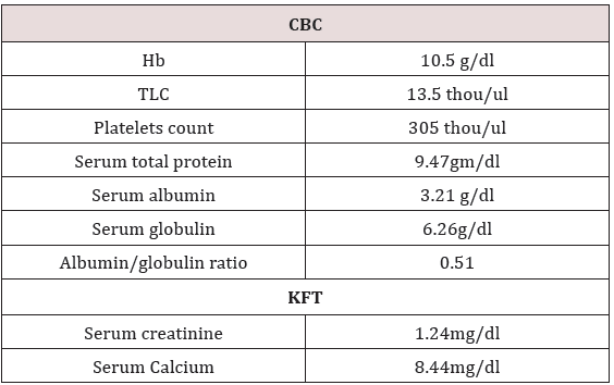
Table 2:
In view of persistent pain in legs MRI dorsal spine was done
which reported well defined altered intensity expansile lesion in
L3 vertebra extending into epidural space. She received palliative
external beam radiotherapy to L3 vertebra 30Gy/10 fractions. She
remained asymptomatic for a month. Then she presented with
complaint of lower limbs weakness and nausea, loss of appetite,
constipation, weight loss (Table 2). USG whole abdomen was done
which reported multiple hypoechoic masses in epigastric region.
Whole body PET CT reported metabolically active soft mass lesion
in gastro hepatic region measuring 9.7x5.5 cm SUV max 20.3, recto
uterine pouch measuring 9.2 x 7.8cm SUV 20.9, left iliac fossa
measuring 7.2x4.0cm- ?mitotic, metabolically active mesenteric
nodes measuring upto 2.2cm-? mitotic and metabolically active
lytic lesion involving L3 vertebra with paravertebral soft tissue
component infiltrating left psoas muscles.
Table 3:
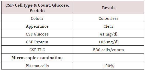
Table 4:
To confirm the diagnosis CT guided core needle biopsy from
supraumbilical mass was done which was compatible with
multiple myeloma. She then received 1st cycle of Bortezomib,
cyclophosphamide and dexamethasone. After few days, she
developed headache, giddiness and syncopal attacks. On evaluation
CEMRI brain reported diffuse leptomeningeal enhancement
involving both cerebral as well as cerebellar hemispheres. Mild
enhancement of basal cistern with associated marginal dilation
of ventricular system (Figure 1). In view of above mentioned
complaints, cerebrospinal fluid study was done (Table 3) (Figure 2).
CEMRI spine was done which reported no abnormal enhancement
in vertebra, cord or thecal sac (Figures 3 & 4). She received 7
fractions (14 Gy) out of 12 planned fractions (24Gy) of whole brain
radiotherapy and two intra thecal injection of methotrexate. MRI
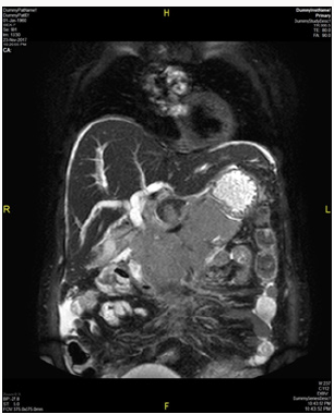
upper abdomen with MRCP reported large mass replacing entire
head and body region of pancreas. Mass encases and compresses
CBD with mild bilobar intra hepatic biliary radical dilation. There
is also encasement of second part of duodenum with possible
infiltrartion of adjacent hepatic parenchyma as well. Features are
suggestive of aggressive pancreatic malignanacy, possibility of
pancreatic lymphoma. Thickening and edema of gall bladder wall
was seen which was suggestive of concomitant cholecystitis. She
was advised for ERCP with SEMS placement which was refused by
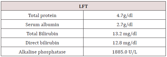
attendants (Figure 5). In view of her deranged liver function test,
both radiotherapy and methotrexate were discontinued and she
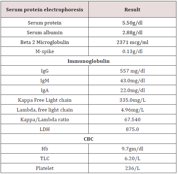
was maintained on supportive care (Table 4).
Figure 1:
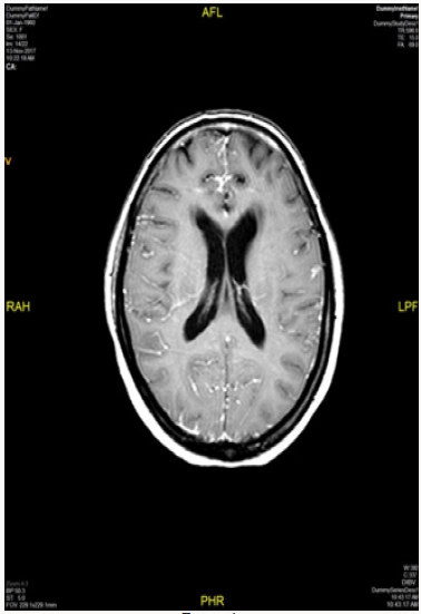
Figure 2:
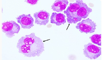
Figure 3:
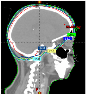
Figure 4:
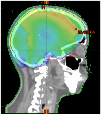
Figure 5:
Discussion
Conclusion
For more Lupine Publishers Open Access Journals Please visit our website:
http://lupinepublishers.us/
For more Open Access Journal of Oncology and Medicine Please Click Here:
https://lupinepublishers.com/Cancer-journal/
To Know More About Open Access Publishers Please Click on Lupine Publishers